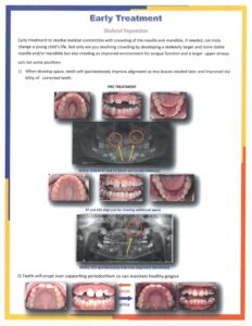
Early Skeletal Expansion Treatment
August 3rd, 2022
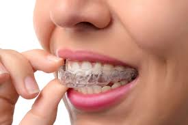
Aligners are an integral tool of orthodontics. Like any tool, when handled properly, it can deliver fantastic results but unlike a hammer, not every patient is a nail. In other words, every patient is different and requires individual and unique treatment planning in order to be successful.
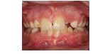
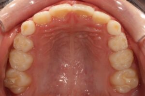
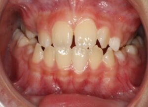
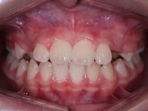
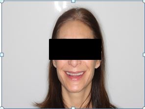
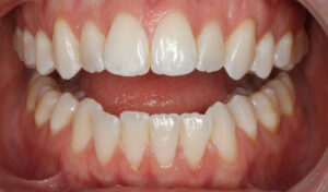
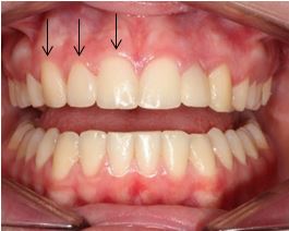
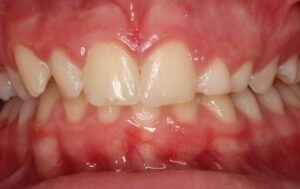
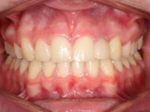
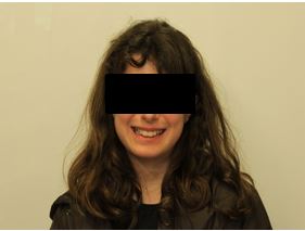
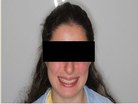
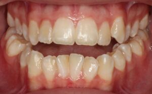
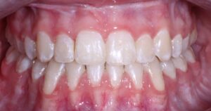
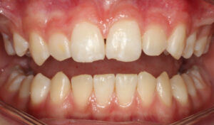
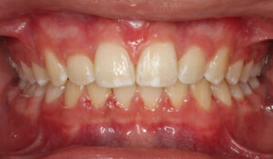
This nice lady provided an example of effective treatment planning to give her a great smile without negatively impacting her fragile gingival health. By using a combination of staged tooth movement. (i.e., moving specific teeth at specific times to avoid excessive expansion and well-designed IPR (interproximal Reduction), we were able to align her teeth for maximum health and esthetics.
IPR is where small amounts of enamel are removed from the sides of the teeth to develop space for alignment to avoid excessive expansion which can compromise the health of the gums.
Her treatment required 8 visits over 14 months
Experience and understanding of the mechanics of the tooth movement are keys to the success with any orthodontics treatment. Let us help.
Preteens Or Teens With Missing Front Teeth
How did we get here?
Do you have a child with a missing upper front tooth? If you do, this blog will provide useful information to help you decide what to do.
There are 3 options.
1) If there is a baby tooth in the position of the missing tooth, keep it . This is not a long term proposition because the baby tooth will be lost (you’ll be lucky if the tooth last 15 years).
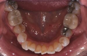
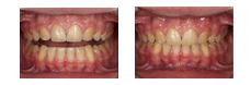
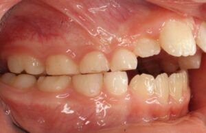
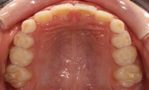
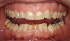
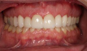
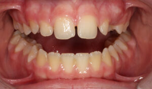
2) Open space for the missing tooth– This is usually the best if you are missing one front tooth, as closing the space for one tooth can create an asymmetry. In the photos below the patient is missing one front tooth. With orthodontics, we created space for the tooth then her dentist placed a Maryland Bridge. 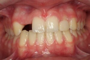

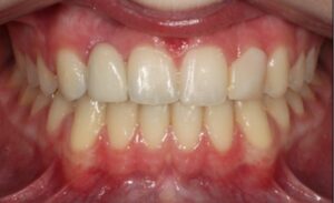
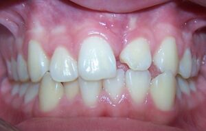
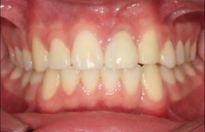
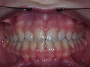
3) Close space for the missing tooth (teeth). This option when possible is the best solution long term as it can alleviate the need for further dental treatment (i.e. the need to replace 2 missing teeth). If handled properly the esthetics of the smile are significantly enhanced. Also you avoid the long term cost for the repeated replacement of the restorations (expect to replace each restoration 4 times over a lifetime). The patient below is a perfect example of this. We closed the space for the two side front teeth and reshaped. 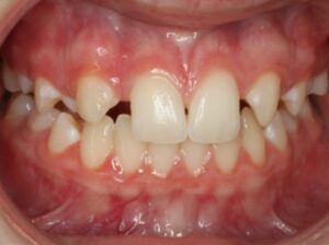
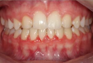
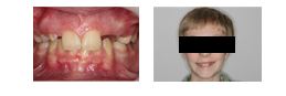
The last example is a patient with a missing front tooth. As I mentioned above, the best results usually involves opening space for the missing tooth because of the asymmetry created but with well planned orthodontics and dental restoration, we were able to develop the esthetics without opening any space.
As you have learned there are options for handling missing front teeth. At our office, we review the possibilities and help you make the right choice for your child.
Let us know how we can help!
10 Things You Didn’t Know About Your Teeth
You use your teeth to bite, chew and talk countless times throughout your day. Unless something is bothersome, you probably don’t give your grill a second thought. So, with our compliments, gnaw on this enlightening list of ten things you didn’t know about your teeth – but guess what – your American Association of Orthodontists member orthodontist did!
Now that you have mastered these tidbits about teeth, you can amaze your friends and family with your trivial knowledge. But your teeth are anything but trivial. Healthy teeth and gums are critical contributors to your overall good health.
Closing Space for Missing Front Teeth
Missing an upper front tooth (or teeth) is difficult for anyone but especially a teen. They have to have a temporary fix ( i.e. Maryland bridge or removable “flipper” with a false tooth on it) until they stop growing. Then an implant (post placed in the bone) can be placed upon which a crown is attached. This can occur in females around 18 years and males 22 yrs. Over the years the crown and implant will have to be replaced (every 15-25 years).
So you can see the process for replacing a missing tooth is long and never ending, not to mention the many times when the cosmetics are just not that good.
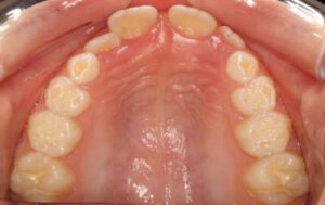
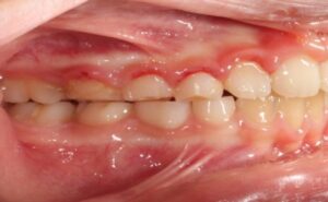
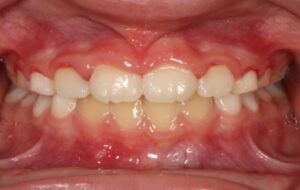
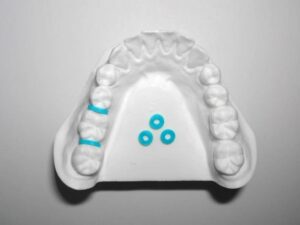
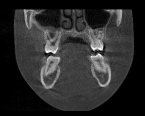
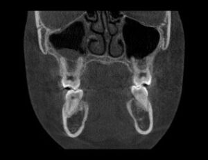
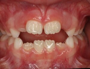
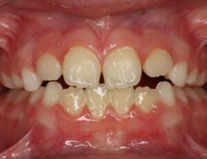
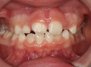
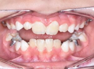
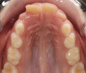
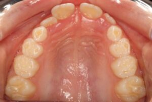
Dr. Michael Sebastian has spent countless hours developing strategies to avoid this potential nightmare by closing the space for a missing tooth. As the following example demonstrates. This 12 year old boy was missing teeth on both sides adjacent to the front teeth (lateral incisors). The strategy for closure is outlined below:
1) Encourage upper eye teeth to come in by the front teeth by selective removal of baby teeth over a 2 to 3 year period. (no braces involved) 4 office visits are needed.
2) Orthodontically manipulate the eye teeth and first bicuspids into a position which simulates the tooth and gum positioning of the teeth they are replacing (lateral incisors and eye teeth).
3) Cosmetically contour these teeth to develop the shape of the missing teeth.
If these strategies are followed then very nice cosmetic results through Orthodontics can be attained. The best of which is they can enjoy their smile when they are teens and don’t have a lifetime of implants and crowns!
Let us know how we can help!
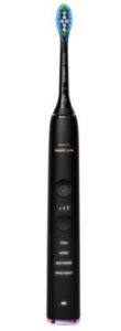
Do you know which electric toothbrush to choose?
There are 2 basic types of electric toothbrushes depending on how the toothbrush bristles rotate.
Well, the American Dental Association recently published an extensive study of these 2 types. Which one was best?
Neither! They both were equally as effective in removing plague thereby improving gum and tooth health (Look Ma, no cavities!)
So, if you are in the market for an electric toothbrush, either of these types will do a great job. Just remember to brush 2 times a day for 2 minutes each time. Focus on brushing the area where the teeth and gums meet. Floss once per day.
Do you clench your teeth when you are feeling stressed? Do you wake up with a headache? Do your jaw muscles hurt? Call our office for an appointment. You may have a habit called bruxism.
WHAT IS BRUXISM?
Bruxism is a habit during which you grind your teeth or clench or thrust your jaw forward over and over again. This habit can affect you’re your oral health. It can cause teeth to break or crack, and increase the chance of gum problems. Adults are not the only ones affected. Studies have found that this can be a problem in children as young as preschool aged.
WHAT CAUSES IT?
Researchers do not know for sure what causes people to do this. Some think stress could be a factor. In preschoolers, studies find an association between grinding their teeth or clenching their jaws and signs of stress such as anxiety or social withdrawal.
Children also can develop this habit when they are losing their baby teeth and their permanent teeth are coming in. Nail biting also may lead to grinding of teeth or jaw clenching. Some children outgrow this, but often adults who grind their teeth or clench their jaws did so as children.
Like many habits, you may be unaware that you do this. You may even do it in your sleep. Tobacco and alcohol use may increase your chances of doing this when you are sleeping. Large amounts of caffeine--for example, 8 or more cups of coffee a day—also can increase this risk. Some medications or illegal drugs may cause users to grind their teeth or clench or thrust their jaws as well.
WHAT CAN YOU DO?
You should contact our office if you notice any of the following:

A number of things can contribute to the problem of bruxism, and there has not been a lot of research on how best to treat it. We may suggest some options, such as:
We also may talk to you about using an oral appliance, which is a plastic tray that fits over either your top or bottom teeth. Use of an appliance may help reduce grinding, clenching, thrusting and may protect your gums and teeth.
Why does my jaw hurt?

Do you have pain in or around your jaws? Does your jaw get stuck? Do you have painful clicking or popping? Are frequent headaches, a problem? If so, you may want to ask us about temporomandibular disorders (TMD).
Your temporomandibular joint (jaw joint) allows you to open or close your mouth and slide your jaw from side to side or back and forth. It is a complicated system of muscles, connective tissues, and the bony joint itself. Because it is so complex, your jaw joint can develop a number of problems.
Some possible causes of TMD include
Diseases that affect the muscles or joints, like arthritis
Other things, like sinus infections, can cause pain in your jaw area. We may want to rule some of these out before identifying TMD as the source of your pain.
DIAGNOSIS
Signs and symptoms of TMD can include
One large, multiyear study also found that people who develop TMD are more likely to report chronic somatic symptoms-like runny nose, fatigue, or dizziness. Anxiety and depression have also been associated with TMD.
If TMD is suspected, we may check your joints and muscles for tenderness and listen for noises like clicking or popping, ask you about pain, and examine how your jaw moves. Symptoms may come and go or may bother you all the time.
TREATMENT
It is difficult to identify the causes of TMD. Treatment usually focuses on relieving the symptoms associated with it.
There are several things you can try that might help:
If your pain is still a problem, we suggest scheduling an appointment for an exam. We might suggest:
CONCLUSION
Because the jaw joint is so complex, it can be difficult to identify what causes pain in and around the joint. Most treatment focuses on relieving the painful symptoms which may be multifactorial and include other specialists. However, most treatments involve reversible modalities which if followed can relieve the TMD discomfort long term!
As we begin 2021, I wanted to express our gratitude for the trust and confidence all of you have shown to our office. You can continue to be confident we are maintaining the high standards of infection control recommended by OSHA and the CDC. My hope, like everyone else is the vaccine proves to be a long term effective method of containing COVID. We are ready to put this in the rear view mirror!
One of the many joys of my practice over the years has been being able to get to know the families we treat. Often mom or dad would come back in the treatment area with their child accompanied by brother or sister. We would have nice conversations. I miss those!
I resolved to pen this letter to let everyone know what I’ve been up to. Last March when we closed the practice for 7 weeks, my wife Tricia and I decided to quarantine at our farm in West Point, Georgia. Our farm is in good shape but needs an assortment of projects accomplished to bring it to the level we hope for. One of the projects on our list was a stone wall between the house and the lighthouse. I decided this was something I could do with the help of our children who would be coming and going from the farm. I thought, this can’t be that difficult? We have an abundance of natural field stone on the property and how hard can it be to stack? Well, little did I know that the project I thought would take only 2 weeks tooth 7 full weeks and over 100 bags of hand mixed concrete! That will be my one and only stone wall project.
At the end of 2019, I had my left hip replaced and 2020 my right. I’m bionic now. I guess my football days caught up with me since no one else in my family has any hip degeneration. The process is straight forward in at 6:00 am, surgery 8:30 walk out at 12:00! I am so thankful we have the opportunity to do this and return to earlier days with no hip pain!
Our family is doing great and it has been nice since two of our children who live in New York City have been able to work remotely so have come home often. We’re all becoming farmers as we have a garden and fruit trees to tend.
I hope all your family is doing well and 2021 is your best year ever!

Do I have to get my wisdom teeth removed after Orthodontic treatment?

The quick answer is maybe, not all patients’ need them removed but some do, the Orthodontist or your Dentist will help you make that decision at the appropriate time.
There used to be a misconception that all wisdom teeth had to be removed after orthodontic treatment because if they weren’t, the teeth would move/relapse. Well, we now know that wisdom teeth do not cause the other teeth to move. There have been many excellent studies which show that whether you have wisdom teeth or not the teeth can move. The best way to assure they don’t is to wear your retainers or if you have a fixed retainer keep it! Just like you brush and hopefully floss your teeth daily to keep your teeth healthy, if you want to keep your teeth straight then 2 to 3 times a week wear your retainer at night.
The reason for wisdom teeth removal includes:
Now, if you have enough space for your wisdom teeth, they are aligned and can be kept clean then keep them.
When is the best time to remove wisdom teeth?
The best time is when the roots are about ½ formed. It is easier to remove these teeth then and there is less chance of having post removal complications like infections, numbness and root fractures. The age range for boys is 16-18y.o. and girls 14-17y.o. It’s not that you can’t remove these at other times, it’s just easier then. Sometimes they have to be removed earlier than this due to their impeding the coming in (eruption) of the second molars.
I hope this helps your understanding of wisdom teeth. Maybe we’ve improved your wisdom!

Are young children self-conscious about their teeth?
The answer is a resounding YES! They may not say they are but if they have crooked teeth or teeth that protrude, new research shows they are self-conscious about it. This research specifically states that they feel less confident about themselves when around others. The study was done on 9 yr. olds who had straight teeth and compared them to those who did not.
So if your child has crooked or protruded teeth and you’re “on the fence” about having them straightened, add improved self-confidence to the list of better oral health as a reason to proceed.

What does a great smile do for a teenager?
As parents, you often wonder if it’s worth it to straighten your child’s teeth. New research has uncovered 3 main themes from interviewing teens post orthodontic treatment:
When you place a value on a great smile, I think these extremely positive benefits as seen through the eyes of teenagers, will increase your support of and confidence in, your decision to have them undergo Orthodontic treatment.

Smart shopping with the ADA Seal of Acceptance
Looking for a dental product to help keep your mouth healthy and your smile bright? The store shelves are stocked with options. How do you know which to try?
How Do I Know Which Products Really Work?
The American Dental Association (ADA) can help. Look for the ADA Seal of Acceptance (Seal) (Figure). The Seal says that a dental product is safe and effective. Also, visit MouthHealthy.org or follow the ADA on social media. The ADA provides information on the science behind dental health claims. Through these outlets the ADA also identifies ideas and trends that are not supported by science. And listen to your dental professionals. They are familiar with your needs and can point you in the right direction.
How Do I Know What A Product Is Supposed To Do?
Products with the Seal make it easy to identify what benefits have been scientifically proven according to Seal program requirements. Companies clearly state, in a bulleted list alongside the Seal, which benefits are supported by science. For example, if a toothpaste says that it helps with cavity prevention, dental sensitivity, enamel erosion, or whitening, the company has provided research showing that it does just that.
And toothpastes are not the only products that carry the Seal. More than 200 over-the-counter products have the Seal, including toothbrushes (both powered and manual), mouth rinses, products that clean between your teeth, and products that help relieve oral pain or dry mouth. The Seal program even has categories for water filters, sports mouth guards and sugar-free chewing gum.
How Do Products Earn The Seal?
Companies cannot use the Seal without permission from the American Dental Association (ADA), and the ADA will not give that permission without sufficient proof that the product does what the manufacture says it will do, safely and effectively.
What qualifies as proof? Science. Companies must provide results from laboratory tests and clinical studies supporting each oral health claim made for a product. These studies have to be performed by laboratories and researchers who are not associated with the company.
The ADA even has a voluntary program to make sure laboratories that conduct independent research in support of products with the ADA Seal of Acceptance can provide reliable results. In addition to reviewing study results and research sites, the ADA conducts its own research on products such as tooth brushes, toothpastes and mouth rinses.
Look For The Seal
The number of dental products in stores and online that make all kinds of promises seems almost limitless. Look for the ADA Seal of Acceptance to be sure that the claims made by the dental product you choose are backed by science.
Tongue Thrust
What is a tongue thrust?
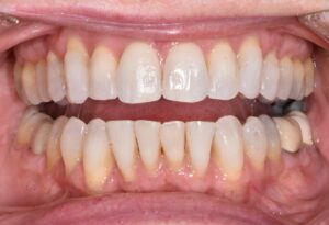
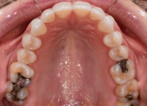
A tongue thrust occurs during swallowing. The tongue should normally move forward and upward compressing against the roof of the mouth to create a seal so swallowing can occur. Instead, it moves forward between the upper and lower front teeth. The pressure every time a swallow occurs exacerbates a front teeth open bite and will often deform the shape of the upper jaw from U-shaped to more V-shaped.

(Pretreatment with front teeth open bite)
 (U Shape) (V Shape)
So, a tongue thrust is functional in that the patient can swallow fine but, it is detrimental to the shape of the upper jaw, the bite and tooth alignment.
Why does a tongue thrust develop?
In many instances, tongue thrusting is the response to a front teeth open bite which was there to begin with. In other words, the patient develops the tongue thrust to compensate for the front teeth open bite. So it is very important in these patients to correct the open bite so the tongue can develop a normal swallowing pattern.
A limited number of people have a tongue thrust without a front teeth open bite. If so, unless there is an associated speech issue then there is no reason to treat. The speech issues are with the “th” and “s” sounds. Have the patient say “there, that, sixty six and sixty seven.” The words will sound slightly slurred, if so, refer to a Speech Therapist.
There are many causative factors for tongue thrusting such as:
-Heredity and thumb or finger sucking are similar since both result in “V” shaped upper arch which makes it impossible for the tongue to fit in the correct position. A specifically designed upper expander with thumb/finger guard works great.
-Enlarged tonsils or adenoids cause the tongue to position forward. This occurs because the upper airway in constricted due to the size of the tonsils so the tongue will have to move forward to open the airway for breathing. Large adenoids make it harder to breathe through the nose so the patient will position the tongue low and forward for airway. The same is true for allergies/nasal congestion. A upper expander with tongue trainer along with removal of the tonsils/adenoids or allergy treatment is the treatment of choice.
-With a tongue tied patient, the lack of tongue mobility makes it impossible for the tongue to move to the correct position. A upper expander with a removal of the frenum is the correct treatment.
-With marcoglossia the size of the tongue prohibits correct positioning. You can’t reduce the size of tongue very often so have to expand the upper jaw as much as possible to accommodate the tongue.
(After treatment with our specially designed upper expander with tongue guard of patient in initial photos)
Now you understand tongue thrust. Let us know if we can be of help!
What it’s like to get an upper jaw expander

Joey needed an upper jaw expander to correct his crowding but he was afraid it would hurt and he wouldn’t be able to eat.
Becky, a team member at the office of Dr. Michael Sebastian could tell Joey was nervous so she made a point of explaining about the expander to help Joey understand what was going on, because you’re usually scared of the unknown.
The first step was to place separators between his back upper teeth (these are small round elastic pieces). She showed them to Joey and explained they would feel like having those teeth flossed when they are placed.
Separators
The separators would stay in until the next week. Becky took Joey’s arm and pressed on it, she asked Joey, “Does this hurt, or does it feel like pressure?” Joey said “it feels like pressure.” Becky assured Joey that the separators would feel like that. She also gave Joey some grape Advil to reduce any possible soreness which might develop.
What a relief! It wasn’t hard at all. Joey could eat whatever he wanted except sticky candy. At Becky’s direction, Joey’s mom would give him some Advil 30 minutes before the next appointment.
Next week, Joey came in to fit for the expander. Becky again was there to make sure Joey had a great experience. As before, she explained what was going to happen that day. They would first remove the separators which had opened a small space for the bands to easily slip on the teeth.

Bands
Like fitting a pair of new shoes, she would have to try on a couple of different bands to get the size perfect. Bands are rings which fit around the two upper six year molars and are attached to the expander. With a gentle touch and Joey’s help, they used a bite stick to push the bands into the correct position.

Bite Stick
With the right bands in position, a scan of Joey’s mouth was done. Becky showed Joey that the scanner is like a very small camera which takes thousands of pictures. You move it slowly around the mouth and on the video screen an exact copy of Joey’s teeth appeared. It only takes about 5 minutes. The orthodontic lab would use this scan and the bands to make Joey’s expander. This was so easy, Joey couldn’t believe how quick and painless it was.
Becky put new separators in so next week the expander could be placed on Thursday at 3:30 pm.
Joey left school for the big day. When he got to the office, Becky gave him Advil. Joey was nervous, it hadn’t hurt so far but he was sure it would today. Becky eased his fear by explaining that today would be just like the last appointment except the two bands they fit would be attached by the expander. They would go through the same process except a special glue would be used to hold the expander in place, nothing like any glue Joey had used before. It was a special glue made for using only in the mouth.
Dr. Sebastian, Becky and Joey worked together to put in the expander. It was just like fitting the bands at the last appointment except for the glue. Joey got to hold “Mr. Thirsty” to remove the flavor of the glue. It took 3 minutes for the glue to set then Becky removed the excess glue and Joey was done! Wow, all the worry and he was fine.

Mr. Thirsty
Becky gave Joey and his mom instructions on care and cleaning of the appliance. She also recommended that Joey take Advil every 4 to 6 hours for the next 24 hours as research has shown that this reduces any discomfort by up to 90%. Becky suggested Joey stay on a soft diet for 5 days until his tongue got used to moving food around the appliance.
Joey and his mom followed these instructions and his first 5 days were nothing like he imagined. He was sore but was able to do all his school work and after school activities. At his next appointment, Dr. Sebastian showed Joey and his mom the change which had occurred over this first 6 weeks, they couldn’t believe it, Joey knew the right thing was being done.


Symptoms of Attention Deficit Hyperactivity Disorder (ADHD) may also be a sign of sleep apnea in children according to experts.
An estimated one to four percent of children experience sleep apnea, according to the American Sleep Apnea Association. Many of the children who deal with this disorder are between the ages of two and eight.
Sleep apnea occurs when a person stops breathing during sleep; it is usually caused by something blocking or clogging the upper airway.
The Centers for Disease Control and Prevention states that around 9.4% of children between 2 and 17 have been diagnosed with ADHD.
Studies show that around 25% of children diagnosed with ADHD may have obstructive sleep apnea (OSA), the American Sleep Apnea Association explained.
Experts say that learning difficulties and behavior issues may be a side effect of "chronic, fragmented sleep."
When sleep apnea occurs, breathing stops during sleep, oxygen levels drop in the body while carbon dioxide levels rise, Norton Children's Hospital explained. This triggers the brain to wake up in order to breathe.
Symptoms of OSA include:
Children who deal with OSA may display the following during the day:
ADHD symptoms include the following:
If you think your child has sleep apnea or ADHD, it’s recommended that parents talk to their child’s pediatrician. At our office, we screen every child for upper airway obstruction which can be a contributing factor to sleep apnea. Ask how we can help!
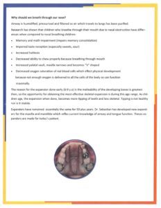
Upper jaw expansion, if needed, is probably the single best treatment you can do for the developing child. The benefits are numerous:
 (Narrow Upper Jaw)
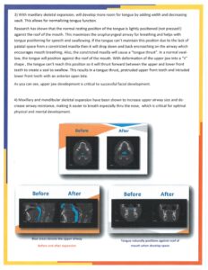
 (Before expansion) (After expansion, airway enlarged)
(Before expansion tongue in low (Normal tongue position after expansion, tongue thrust position) tongue in roof of mouth)
(Before expansion and tongue thrust (After expansion and resolution of treatment) tongue thrust)
All this, and there is no mention yet of the skeletal and dental improvements which are achieved with upper jaw expansion.
 (Before) (After)

 (Before expansion) (After)
Best Age?
We’ve covered all the benefits of upper jaw expansion. Now let’s understand when is the best age for expansion. Research has shown the best time is between the ages of 6 and 10 if you want to maximize the above benefits plus increase the stability. The upper jaw is very malleable during this stage of development so if expanded properly the upper jaw responds well and the results are stable, long term.
This type of expansion is skeletal, the other type of expansion is dental, this is when the teeth are tipped out which is not stable. Prior to age 10 expansion is about 85% skeletal 15% dental, as the child ages the numbers reverse. By 17 y.o. the ability to get skeletal almost zero.
Type of Expander?
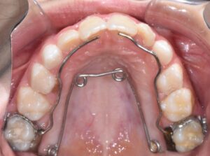
The last area of concern is the type of expander used. Again we rely on good research to experience to choose the expander. The fixed quad helix is the top expander for results achieved. We have been using a specifically designed fixed quad-helix for 20 plus years. We custom design the appliance for not only posterior expansion but also anterior expansion, tongue restraining in tongue thrusting patients and crib for finger sucking. By doing this we don’t have to use different appliances and can treat multiple areas with one appliance. The expansion requires 6-8 months with 6 months retention for long term stability.
Don’t hesitate to contact us if we can be of any help!

What does Chlorine do to your Teeth?
Often times we have patients with discolored enamel around their brackets. One of the first things to take in consideration, especially in teens, is every day activities, like swimming.
Chlorine is one of the leading causes of teeth discoloration and many people aren’t aware of the problem because you can’t see the pH balance of a pool. This is also known as “Swimmer’s calculus.” Pools have a high pH level that stains teeth brown and prevents saliva from doing its job in cleansing the mouth. Poor pH balance in a pool can also cause the enamel of teeth to soften, making teeth more susceptible to damage and decay, as well as more sensitive in general.
As the enamel of your teeth wears down from exposure to chlorine, it becomes discolored. Poor enamel health is common in competitive swimmers because of prolonged exposure to chlorine which could also cause sensitivity.
How can you protect your teeth from Chlorine?
The best recommendation to prevent chlorine from staining your teeth, would be to try and keep your mouth shut while you’re in the pool. This is nearly impossible to prevent, but the less amount of pool water you get in your mouth, the better.
Another important thing to do, is to brush and floss your teeth as soon as you get out of the pool. You don’t want the chlorine to sit on your teeth any longer than necessary. We suggest always bringing your toothbrush when you go to the pool to swim.
If you love to swim, make sure you have toothpaste with MI Paste or baking soda. Both of these can help fight the acid caused by pools and lessening the chances of chlorine discoloration of your teeth.
Can my child benefit from an Orthodontic Expander?
This is a common question concerned parents often ask. They also want to know if their child needs an expander then when is the best age to do it. In this blog we will cover these questions.

Upper Expander
When parents talk about expanders they are usually referring to upper jaw expanders (maxillary expansion). The lower jaw can also be expanded but that is a topic for another blog. We will focus on upper expanders here.
Let’s answer the age question first. The best time to do an upper expander is between the ages of 6 to 10 years old. The reason for this is the bones through the sutures are very malleable so are easily reshaped. The older a patient gets the harder the bones are to reshape and the less stable they are when you do.
Narrow “V” shaped upper jaw
There are two types of expansion
1. Dental
2.Skeletal
The best type of expansion in most instances is skeletal which is where the bones are reshaped because the results are more stable and the teeth are positioned more ideally over the bone which is healthier. So if you want the best expansion as mentioned earlier then do it in the 6-10 year old range. Dental expansion is where the bone stays the same shape and the back teeth are pushed out. This works best when the back teeth are angled inward.
Now, what are the benefits of using an Expander?
Pre expansion Post expansion
Pre 3D expansion Post 3D expansion
The key is to have a properly designed expander which can function in 3D. They have to be fixed (removable expanders, ones that can be taken in and out, do not predictably work!) They also should not have slots which need to be turned by you with a key. The slot is usually turned every other day which causes discomfort and is difficult to do.
Our office specifically designs each fixed expander to fit the needs of each child. Our patented 3D expander can work in all directions to achieve the most effective results. Other expanders only work in one direction. We activate the expander every 6 weeks so instead of potentially being uncomfortable every other day, the discomfort would be every 6-8 weeks for 3 to 4 visits. The 3D expander stays in for 6 months followed by 6 months of retention to ensure stability.
What are the downsides of expansion?
Really none, the predictability, the reduced need for extractions, the stability, less time in braces as a teen and better results! Plus as we mentioned it can help with sleep and optimum facial development.

If your child is in the 6-10 year old range, call us for a complimentary consultation.
Do vibrating or light emitting devices move teeth faster?
I am often asked this question on new devices that promise faster movement of the teeth which should result in shorter treatment times.
The current unbiased research (in other words, research that is not paid for by the manufacturer of these devices!) doesn’t support any claims of shorter treatment times. So if you have a friend who claims their teeth are moving faster because of using one of these devices or a dentist or orthodontist claims they will accelerate treatment, the research does not back up any of these claims. If it did you can be sure we would use them at Elite Smiles plus are there any long term (haven’t studied) negative consequences of using them, we don’t know.
Call or email if you’re ready to “build a better smile”

Smile Direct Club
If you watch tv or are on social media then you have to be aware of Smile Direct Club. It is a method of moving teeth without direct supervision of an Orthodontist. I can tell you as someone who has done thousands of patients using doctor monitored Invisalign that direct supervision is critical to the success of treatment.
As has been anticipated by Orthodontists, Smile Direct Club has passed 1,000 complaints on the Better Business Bureau’s website in a short time. I personally, have 2 patients whose bites (the fit of their teeth) were significantly altered for the worse by using Smile Direct Club. They came to us after using Smile Direct Club when they realized their bites were “way off” and could no longer chew properly. Both now have to undergo comprehensive orthodontic treatment with braces for up to 2 years!
If you or anyone you know is going to consider this route to straighten their teeth, at least visit an Orthodontist prior to treatment to discuss what your options are.
Orthodontics and your Family

Why should I choose Dr. Sebastian as my Family’s Orthodontist?
A pioneer in early orthodontic evaluation and treatment of the developing child, Dr. Michael Sebastian has the experience and expertise to critically assess the growing child’s mouth, upper airway and surrounding facial structures. His timely holistic approach is designed to encourage optimum facial esthetics supported by a healthy, stable and beautiful smile.
The first visit with Dr. Sebastian is recommended at 7 years old. This appointment includes a health, dental and airway questionnaire review, a comprehensive photographic examination, a 3 dimensional radiographic analysis along with a thorough orthodontic exam, upper airway and facial assessment all accomplished in a welcoming comfortable child friendly environment.
Dr. Sebastian will summarize all the findings in a priority format so you will know what and when, if anything, needs to be done for your child.
There are two basic directions which can be recommended:
12 years old
All the permanent teeth are usually in at 12 years old. At this time, your child is evaluated for braces or Invisalign to finalize the alignment, fit and esthetics of their teeth. Now, not all children are seen on this schedule as their dentist may not have referred them at 7 years old. They may follow your child until all the permanent teeth are in then make the referral around 12 years old.
If my child doesn’t see Dr. Sebastian at 7 years old can they still be helped? Yes, Dr. Sebastian will provide a complete assessment with recommendations at any age.
My child is almost a teen, what can be done? Treating teens is the original reason Dr. Sebastian became an Orthodontist, he enjoys interacting with and understands how to work with teens.
There are 2 keys to successful orthodontic treatment of a teen:
Experience, communication, comfort, choice and excellence are the principals by which Dr. Sebastian connects with every one of his young patients. You can feel secure in your decision to entrust us with your child.
If you’re 50 plus years old, is it ok to undergo Orthodontic treatment?
Many adults want to improve the appearance and health of their teeth but are concerned about the length of time and possible negative consequences of undergoing Invisalign or conventional orthodontic braces.
Well, more and more research is showing that adults do just as well with orthodontic treatment as teenagers even if the adult has compromised gum disease as long as it is under control.
Yes, the research presents clear evidence that not only do adults get the straight healthy teeth they’re looking for but also the length of treatment and possible negative consequences (ex. increased TMJ or gum disease) are no different for adults than teens!
Call or email us if you have questions or are ready to do something about your smile. Don’t be saying “I wish I had done something earlier”

Do you have sensitivity of your teeth at the gumline?
This is often due to gum recession which exposes an area of the tooth or teeth which is not meant to be exposed to the mouth. This area is covered by a thin layer of tooth (cementum) which is easily worn away with tooth brushing. As a result the tooth will become sensitive to almost anything which touches it.
If this is happening in your mouth the first thing you should do is:

What are the elements of a great smile?
Dr. Michael Sebastian
We all know when we see a person smile that “wow” he or she has a great smile!

But do we know what makes up a great smile? What are the elements of a fantastic smile? Today we will discuss and break down these elements for your understanding.
The smile arch. This esthetic ideal means the upper front six teeth should form an arch which parallels the lower lip. A reverse smile arch or flat smile arch is not pleasing to the eye.

Notice upper front teeth form arch which parallels lower lip
Flat smile arch
Reverse smile arch
2. Show all the upper front teeth with a normal or relaxed smile.
3. The width of the smile- With a relaxed smile, the upper back teeth should be visible and no dark spaces between them and the cheeks should be present.
4. The upper midline (which is between the upper front teeth) should be centered in the philtrum of the upper lip.
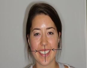
This smile shows elements 2, 3 and 4
Don’t use the nose because the nose is often deviated from the facial midline. Studies have shown the upper midline can deviate 3mm from the centered position before it becomes unaesthetic. The upper midline doesn’t have to match the lower midline for esthetics.

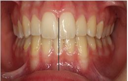
Missing lower front tooth so upper and lower midlines can’t match
5. The proportions of the teeth both in length and width need to match each other and the size of the face. The upper front teeth are the widest, the teeth next to those are ¾ their width and the eye teeth are 9/10 their width.
6. The color should be light and even throughout the front teeth.

Front teeth are short due to wear. Color is too dark

Now teeth are normal width, length and color after treatment

Small teeth

After treatment to align and restore teeth to correct size
7. The gum levels should be as noted in photo with the front teeth gum level higher than the side front teeth by 1-2mm and the eye teeth the same height as the front teeth.

Gum levels not correct

After treatment to develop correct height of gums and width of teeth
8. The upper front teeth don’t overly protrude or retrude. They should position just in front of the lower front teeth with the teeth together.
Before
After treatment to correct protruded upper front teeth

Retruded upper front teeth

After treatment to correct upper front teeth retrusion
9. The edges of the teeth should be smooth
10. The gums should fill out the space between the teeth. No what we call “black triangles”.
Rough edges and black triangle
After treatment to align, correct black triangle and polish rough edges
Now, when you add all these elements up you get a fantastic smile which radiates confidence, beauty and health.
Let us “Build You” a better smile
I am asked Often about the use of Baking Soda toothpastes. Should they be used, are the safe, are they effective and are they better or worse than Other toothpastes. In today's blog we'll answer some of those questions.
What is baking soda toothpaste? It is toothpaste with the primary active ingredient of sodium bicarbonate.
Are they safe?
Yes, extensive research has Shown toothpastes containing baking soda to be safe. An interesting side note is patients swallow 5-7% of the toothpaste When brushing and there are no negative effects here either even for patients on a low salt diet.
Are they effective?
Yes, they are effective at killing the bacteria which causes cavities. Patients who use these toothpastes have improved oral health including the teeth and gums.
Are they better or Worse than Other toothpastes?
The best reason to use baking soda toothpastes iS because Of their IOW abrasiveness and improved tooth Whitening. Most toothpastes now are very abrasive. Abrasiveness is good to clean Stain and Whiten but it also causes erosion Of the teeth. so if you are prone to erosion of your teeth then Baking Soda toothpaste is an excellent alternative because it also is excellent at whitening.
So yes, I Can recommend Baking Soda toothpaste because it is safe, kills the bugs,
reduces erosion and whitens.
Do I have to wait for the Dentist to send my child to the Orthodontist?
The simple answer to this question is NO. If you notice the permanent teeth are coming in crowded, with excessive space, the front teeth are protruded or retruded, the back or front upper teeth bite inside the lower teeth then you need to take your child to an Orthodontist.


You may assume the dentist is aware of these problems and that they will make the referral to an orthodontist but sometimes this does not occur. Dentists are often focused on the health of the teeth and gums. They look at how good the child’s brushing and flossing is and if there are cavities. They don’t often concentrate on how the teeth fit together (the bite). This is what Orthodontists specialize in doing.
The American Association of Orthodontics recommends a consultation with an Orthodontist at age 7. As most poor bites start at this age. By examining a child at this age, the Orthodontist can diagnose many potentially damaging bite and jaw development conditions before they cause major problems. If treatment is necessary at this critical stage, then results can be dramatic.


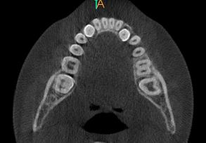
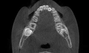
The other reason for an examination of children at this age is the detection of Sleep Disorder Breathing or SDB. It is often the precursor to Sleep Apnea. Using our diagnostic criteria and ultra-low dose CBCT radiography, we can learn if a child has an upper airway disorder (think narrow airway) which could affect their ability to get enough oxygen so their brains and bodies can develop to their maximum potential. We use this radiograph for our orthodontic purposes but can also use it for airway analysis so the child gets the additional benefit without any additional radiation! As parents, we all want to give our children the best chance for success and without a big upper airway that is not possible.
As you can see, there are many benefits to examining a child early. Not all children will need orthodontic treatment at this stage but you don’t want to miss the opportunity this age of development offers. You don’t want to be saying “I wish we had of”
At the orthodontic office of Dr. Michael Sebastian, we are here to guide your child and family through this critical stage of development.
Are thumb and finger appliances necessary?

Protruded upper front teeth, can easily be damaged in accidents plus the child can suffer from social teasing. Crossbites of the back teeth, can cause excessive wear and fracture of the permanent molars.
If the habit is stopped prior to 3 years old the deformation will usually reverse itself without orthodontic intervention. A sequence of treatment is recommended for the greatest success. First-Talk with your child, let them know that their habit is “hurting their teeth.” Set up a calendar of positive reinforcement (prizes can help meet goals) For example, 2 days without the habit, 5 days without, 10 days, 20 days, 40 days, then 60 days. After that, it is rare to get relapse. I know you’re going to say, “but my child can’t count to 60,” that’s the reason you set up a calendar in a prominent place like the refrigerator. Have them place a mark on the calendar for each day of success. Pre-pick a prize for each goal reached.
If you can tell your child is trying, but not successful then you can try “Stops it,” (make sure no allergies!) from the pharmacy. This when put on the offending digit, gives it a bitter taste which acts as a reminder not to put the finger in the mouth.
Second-If not successful in 2 months with the above protocol, then an orthodontist can help. At our office, I repeat the above regimen with new additions as an outside authority figure, that may be all that is needed to stop the habit. If not successful in 2 months, then we proceed to the fixed (not removable) upper guard.

I find if we try the “benefit method” first then the child is much more accepting of a fixed guard. The appliance is left in for 6 months.
What if the child’s upper jaw has been permanently deformed? At the office of Dr. Michael Sebastian, we combine a thumb/finger guard with an upper expander to not only resolve the habit, but also to correct the skeletal deformation which occurs.


The last thing to consider is a tongue thrust (October 10/2018 Blog). Often, a tongue thrust will result after a thumb or finger habit deforms the upper jaw so even after the habit is resolved the tongue thrust will continue to deform the upper jaw. That is why is imperative to treat the tongue thrust while treating the upper jaw deformity. We have developed a special upper expander which treats all of these considerations.


At the office of Dr. Michael Sebastian, we emphasize the treatment of thumb and finger habits for successful development of the teeth and face.
Clear aligners, Invisible braces, Invisalign

Clear aligners, Invisible braces, Invisalign (a brand name that has become a generic term like Coke even though many companies make clear aligners) are now a part of the Orthodontic tool box used to move teeth to build healthy beautiful smiles.
Like braces clear aligners are designed to move teeth in small increments. Also like braces, aligners need to be planned in a specific sequence in order to move the teeth most efficiently into the correct position. “You can’t move all the teeth at once”. Most importantly, aligners are not a “do it yourself “ technique, the computer technician moves the teeth under the guidance of the Orthodontist. The Orthodontist, not the technician, has the education, experience and skill to move the teeth correctly within biologic limits to ensure the health of the teeth, surrounding gums and jaws.
Once a plan is arrived at, the trays are fabricated to make sequential movements through multiple trays with each tray designed to make specific tooth movements. With each tray, specific teeth are reset into a new position and the tray is fabricated. Over the course of wearing the tray 7-10 days the teeth are gently guided into the desired next step. The trays must be worn 21-22 hours per day to be effective. It’s not like if you wear them less than the desired time the teeth still move but at a slower pace, they don’t move at all! So effectively, the only time they aren’t worn is for eating, drinking and oral hygiene,
The trays are made of a pliable polymer which gently flexes to put pressure on the teeth to be moved. Sometimes this is not enough to effectively move a tooth, so small tooth colored attachments are added to the outside of a tooth to enhance the desired movement. The total number of aligners needed depends on the complexity of the movement.
Advantages of clear aligners include:
Disadvantages:
Clear aligners can be used to treat teens as well as adults. In fact, the oral hygiene is much better in teen clear aligners than braces. As long as the teen doesn’t try to drink beverages containing sugar, such as soda, fruit juices and sports drinks with their trays in, then the oral hygiene is never a problem with clear aligners which is a plus.
Braces versus clear aligners is a choice which you should not make alone. We are here to help you at the orthodontic office of Dr. Michael Sebastian.
Questions to consider when thinking about using online teeth straightening services
I don’t know if you have seen all the advertisements popping up from “Do it yourself” teeth straightening companies like Smile Direct Club, Candid Co., Smile Love, SnapCorect, Orthly, etc.
If you know of someone who is considering using this route to align their teeth, then you might offer these questions for them to ask prior to starting treatment. These questions were developed by the American Association of Orthodontics to help consumers make a well informed decision.
YES
NO - Are you comfortable starting orthodontic treatment without comprehensive diagnostic records? If you still want comprehensive diagnostic records taken, are you alright going to another dental professional to take them? If yes, what will that cost?
YES
NO - Are you comfortable with orthodontic treatment that does not involve any in-person visits with a dentist or orthodontist?
YES - How do you know that is the best treatment option for you, given your unique situation and oral condition, compared to other treatment models (such as braces)?
NO - How is the decision being made for the best treatment model for you, and who is making that decision?
YES
NO - Are you comfortable not being able to research your dentist’s or orthodontist’s background, credentials, patient reviews, etc. before you begin treatment?
These are some of the questions which any consumer should ask prior to starting treatment. Moving teeth is not a simple process and requires the oversight of a skilled dental professional.
Stun Your Readers
“Be original, show off your style, and tell your story.”
“My child, Cody has always slept with his mouth open and snored. I remember sitting in his room watching him struggle to get a good night sleep because he couldn’t breathe properly. He started his treatment with Dr. Sebastian in October of 2013. Within months, I saw a change! Even during the day, I would notice he was keeping his mouth closed to breathe and by the time his treatment was finished, he had completely stopped snoring!! It has been amazing to see such a simple appliance make such a huge difference in the life of my child!! Thank you Dr. Sebastian for your dedication to each child you treat.”
As you can see from this testimonial, the importance of early treatment is imperative. Utilizing this technology, we are convinced that orthodontics must be more than just about a healthy bite. We continue to use our sleep questionnaire and visual examination of the soft tissue skeletal appearance, ability to breathe through nose and tonsil size. We have also begun a tracking system that will allow us to better follow the progress of our Phase I patients who are experiencing any airway problems.
80% of symptomatic airway children go un-diagnosed, so the problem is not going to just “go away” on its own. Dentists and orthodontist should be at the forefront of this effort because we are the ones seeing these children on a regular basis and it fits into areas in which we are intimately involved. Our goal is to help the next generation “breathe a little easier”.
As always, we appreciate your continued support and trust in our office. If I can answer any questions you may have, please do not hesitate to contact our office.
Orthodontics - April 30, 2014 - Vol. 28 - No. 5
After clinical crown lengthening, endodontic therapy, and prosthodontic treatment, teeth have a survival rate of 96% at 5 years and 83% at 10 years.
Article Reviewed: Long-Term Survival Rate of Teeth Receiving Multidisciplinary Endodontic, Periodontal and Prosthodontic Treatments. Moghaddam AS, Radafshar G, et al: J Oral Rehabil; 2014;41 (March): 236-242.
Background: With larger numbers of adult patients now seeking orthodontic treatment, orthodontists are frequently involved in interdisciplinary treatment planning regarding compromised teeth.
Objective: To evaluate the long-term survival rate of teeth undergoing endodontic, periodontic, and prosthodontic treatment.
Design: Retrospective study.
Participants/Methods: 87 patients (81% female; age range, 21 to 70 years) who underwent crown lengthening, endodontic treatment, and prosthodontic work on at least 1 tooth between 1996 and 2009 at the Guilan University of Medical Sciences were included. A total of 245 teeth were treated. Teeth with furcation involvement, considerable mobility prior to crown lengthening, or a crown-to-root (C/R) ratio <1 were excluded. All crown lengthening procedures were done by a single periodontist whose records were used to select the sample. Patients were recalled for a clinical and radiographic exam to record bleeding points index (BPI), position of the restorative margin relative to the gingival margin, pocket depth, mobility, C/R ratio, and reasons for any lost teeth. Teeth with severe caries requiring addition crown lengthening, extensive periodontal lesions, pocket depths >7 mm, or severe furcation involvement were deemed hopeless.
Results: 18 teeth (13 maxillary, 5 mandibular were lost or deemed hopeless during the recall exam. The survival rate was 98% for 3 years, 96% for 5 years, 83% for 10 years, and an estimated 52% for 13 years (using the Kaplan-Meier estimator). Survival rate was not influenced by patient sex, history of smoking, or the presence of a post. Teeth that had survived >10 years showed increased pocket depths and C/R ratios. When examining factors to predict failure, the major determinates were found to be C/R ratio and the position of the crown margin relative to the gingival margin.
Conclusions: The survival rate of teeth receiving complex prosthodontic, endodontic, and periodontic treatment was 83% at 10 years.
Reviewer's Comments: The authors highlight the fact that these survival rates reflect good interdisciplinary treatment planning, and did not attempt complex treatment if the tooth was overly compromised. It also was unclear what the response rate was for patients being recalled, which could alter the strength of these findings.(Reviewer–Brent E. Larson, DDS, MS).
© 2014, Oakstone Publishing, LLC
Orthodontics - April 30, 2014 - Vol. 28 - No. 5
The discontinuation of excessive gum chewing can effectively eliminate chronic headaches in some adolescents.
Article Reviewed: The Influence of Excessive Chewing Gum Use on Headache Frequency and Severity Among Adolescents. Watemberg N, Matar M et al: Pediatr Neurol; 2014;50 (January): 69-72.
Background: It is not uncommon for adolescents to suffer from chronic migraine or tension headaches. Is there something that we might be able to do to help them?
Objective: To assess the impact of excessive gum chewing on headache occurrence among children and adolescents.
Participants: 30 subjects who reported chronic headaches and excessive gum chewing.
Methods: The sample was divided into 4 groups based on the amount of gum chewing per day. Group 1 was up to 1 hour; group 2, 1 to 3 hours; group 3, 3 to 6 hours; and group 4 >6 hours per day. All of the patients were asked to stop gum chewing for 1 month. At that point, their symptoms were evaluated and they were asked to renew their gum chewing habit exactly as it was before discontinuation; a second interview was carried out 2 to 4 weeks later.
Results: Following the discontinuation of gum chewing, 19 of the 30 patients reported complete resolution of headaches and 7 described some improvement in headache frequency and intensity. No improvement occurred in 4 patients. The duration of the headache symptoms before stopping gum chewing did not play a role in the clinical response because some children who reported full or significant improvement had suffered from chronic headache for up to 6 years. All 20 of the patients who reported either complete or partial headache relief reported relapse of their headaches within days to a week of resuming gum chewing. Ten of the 30 patients in this study reported chronic symptoms related to the temporomandibular joint, and these symptoms also improved upon gum chewing discontinuation.
Conclusions: The discontinuation of excessive gum chewing can effectively eliminate chronic headaches in some adolescents.
Reviewer's Comments: This is a clean-cut and impressive study that provides an opportunity to provide a significant service to our patients. Imagine being able to help a patient who has suffered from chronic headaches for 6 years to eliminate these symptoms by simply discontinuing gum chewing.(Reviewer–John S. Casko, DDS, MS, PhD).
© 2014, Oakstone Publishing, LLC
Orthodontics - April 30, 2014 - Vol. 28 - No. 5
The likelihood of having tinnitus was 8 times higher in individuals with temporomandibular disorder (TMD) symptoms than it was in those without TMD symptoms.
Article Reviewed: Is There a Link Between Tinnitus and Temporomandibular Disorders? Buergers R, Kleinjung T, et al: J Prosthet Dent; 2014;111 (March): 222-227.
Background: Patients who have temporomandibular joint and masticatory muscle disorders (TMD) also are reported to frequently have tinnitus. Reports range from 2% to 59% of TMD patients also having tinnitus. Are TMD and tinnitus related?
Objective: To investigate if there is an association between tinnitus and TMD, and to examine if therapy for TMD has an effect on tinnitus symptoms.
Design/Methods: This was a prospective clinical study in which the prevalence of TMD and tinnitus was investigated in 951 consecutively referred prosthetic patients with a mean age of 54 years. Thirty patients with both TMD and tinnitus were the test sample examined in the study. A baseline examination was performed on each symptomatic patient by one experienced dentist. The examination consisted of a functional analysis of the masticatory system, an evaluation of the temporomandibular joint and associated musculature, and a tinnitus questionnaire. Each patient received dental functional therapy, which consisted of physiotherapy and intra-oral splints. The patients were evaluated at 3 to 5 months to determine the effects of functional therapy on the TMD and tinnitus symptoms.
Results: 8.6% of the 951 subjects were diagnosed with TMD and 7.2% were diagnosed with tinnitus. In total, 3.2% had both TMD and tinnitus simultaneously. The likelihood of having tinnitus was 8 times higher in individuals with TMD symptoms than in those without TMD symptoms. Eight patients had unilateral TMD symptoms and unilateral tinnitus; all were on the same side. Stomatognathic TMD therapy improved tinnitus symptoms in 44% of the test subjects.
Conclusions: This trial showed a significant correlation between tinnitus and TMD. Dental functional TMD therapy may have a positive effect on subjects with simultaneous symptoms of both TMD and tinnitus.
Reviewer's Comments: Previous studies and this report appear to support a connection between tinnitus and TMD. With an incidence of tinnitus that is 8 times higher in patients with TMD symptoms, perhaps a question about tinnitus in addition to TMD would be appropriate in our patient health history forms. A significant placebo treatment effect has been reported in TMD and tinnitus patients, and I agree with the authors that it would be interesting to assess the TMD/tinnitus treatment effects with a longer-term randomized trial including controls.(Reviewer–John S. Kanyusik, DDS, MSD).
© 2014, Oakstone Publishing, LLC
Orthodontics - April 30, 2014 - Vol. 28 - No. 5
You can use this article as an objective basis to advise your patients that they will not be seen as any less attractive by others as a result of wearing braces.
Article Reviewed: Impact of Metal and Ceramic Fixed Orthodontic Appliances on Judgments of Beauty and Other Face-Related Attributes. Fonseca LM, de Araújo TM, et al: Am J Orthod Dentofacial Orthop; 2014;145 (February): 203-206.
Background: Patients are sometimes reluctant to pursue orthodontic treatment because they feel that wearing braces will result in them being seen as less attractive. Is there a basis for this concern?
Objective: To investigate how people who wear fixed orthodontic appliances see themselves and how they are seen by others in social settings.
Participants: 60 adults (21 men, 39 women; ages 18 to 47 years) whose maxillary dentition was complete with both dental arches either aligned or having mild crowding.
Methods: 3 smile photographs were taken for each subject: one with metal braces and an 0.018" stainless steel maxillary archwire, a second with ceramic braces and a maxillary 0.018" stainless steel archwire, and a third acted as a control with no braces in place. The subjects rated each of their 3 photographs on an analog scale ranging from 0 to 10 from not beautiful at all to very beautiful. Additionally, the same 180 photographs (3 each of the 60 subjects) were rated by 15 adult raters who were lay people not currently undergoing orthodontic treatment.
Results: The subjects saw themselves as more beautiful when not wearing a fixed orthodontic appliance, followed by wearing an esthetic fixed orthodontic appliance, and finally a metal fixed orthodontic appliance. However, for the raters there was no statistically significant difference found between any of the 3 photographs.
Conclusions: Wearing an orthodontic appliance has no bearing on interpersonal esthetic judgments.
Reviewer's Comments: There is no doubt that there are people who do not pursue orthodontic treatment because they feel that wearing braces will make them look less attractive. I suspect that many orthodontists tell their patients that they understand the concern about looking less attractive wearing braces; however, people looking at them will not. Now you have a study to provide a basis for that statement, and I would suggest that you might even want to keep a copy of this article in your office.(Reviewer–John S. Casko, DDS, MS, PhD).
© 2014, Oakstone Publishing, LLC
Dr. Powell is currently accepting new patients and taking appointments in April for West Wieuca. To make an appointment for your child, please call 770-934-5900.
Dr. Wesley Powell graduated from the McCallie School in Chattanooga, Tennessee. He then attended Southern Methodist University in Dallas, Texas, where he earned a degree in Anthropology. He attended Columbia University School of Dentistry and Oral Surgery in New York, New York for his dental training. Dr. Powell then completed a three-year residency program in Pediatric Dentistry while earning a Masters Degree in Clinical Dentistry at the University of Alabama at Birmingham.
Upon completion of his residency, Dr. Powell practiced for two years in suburban Washington, D.C. at a pediatric/orthodontic practice before relocating to Atlanta. While there, he was on staff at Arlington Hospital.
Dr. Powell is a member of the American Academy of Pediatric Dentistry, Southeastern Society of Pediatric Dentistry, American Dental Association, Virginia Dental Association, Georgia Dental Association and the Better Business Bureau. Dr. Powell served as the Atlanta representative for Crest, Proctor and Gamble for Children’s Dental awareness month speaking to inner city children and also appeared on local television to promote dental health. He is currently a member of Gnathos, a continuing education organization that deals solely with the orthodontic treatment and clinical modification of growth and development of growing children and teens, Atlanta Dental Coordinator for the charitable organization Team Smile and the Atlanta Falcons. This organization teams with a local professional sports team to provide free dental care and screenings to children in many NFL/NBA cities including some colleges (including UGA!) around the country.
Dr. Powell resides in Buckhead. He has one son, Pearson, and two Jack Russells Sophie & Cowboy. He enjoys golf, travel, snow skiing, the outdoors and fishing. He is active in the community with numerous foundations and community service organizations including the National Black Arts Festival, Camp Sunshine, and Lanier Partners of North Georgia. Dr. Powell is a member of Peachtree Presbyterian Church and has participated with helping with pre-school children.
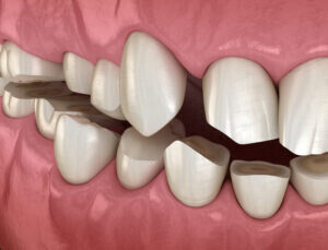
The greater the space and the more mesially located the lateral spacing was, the more unattractive the smile.
Background: Facial esthetics and the attractiveness of a smile are of great interest to clinicians. Previous research has described the negative impact of having a maxillary midline diastema on perceived esthetics.
Objective: To evaluate the perception of smile esthetics among laypeople and orthodontists as affected by the presence of diastemas in the maxillary lateral incisors using an oblique smile analysis.
Methods: The study used 2 standardized oblique smiling photos from 2 female subjects. One subject had been treated with extractions and one without extractions, and both were considered to have attractive smiles. The photographs were digitally altered to create interproximal spaces in the lateral incisor. Space were generated in 0.5mm increments and were located in the mesial, distal, or both surfaces. The final images were randomly assembled and given to 120 judges for evaluation. Sixty of the judges were orthodontists and 60 were laypersons. The judges were asked to assess the attractiveness of the images on a visual analog scale.
Results: the judges rated the images without lateral spacing as the most attractive smiles. both groups rated the presence of diastemas as unattractive. they found that the greater the space and the more mesially located the lateral spacing was, the more unattractive was the smile rating.
Conclusions: This study suggested that spacing in the upper lateral incisor area is a factor in smile attractiveness. The larger and the more mesially located a lateral diastema, the more unattractive was the smile assessment.
Reviewer's Comments: This was an interesting article in that the focus was on the esthetics of lateral spacing rather than the more commonly studied midline diastema. Both laypersons' and orthodontists' perceptions of the lateral spacing were quite similar; the larger the space and the closer to the maxillary midline the less attractive the smile. For the layperson group, a 0.5mm space distal to the lateral was not rated as unattractive. thus, if it is not necessary to leave some space in the maxillary anterior arch, this study supports leaving the space distal to the laterals. There were no differences found between the extraction smile photos and the non extraction smile photos. (Reviewer-John S. Kanyusik, DDS, MSD)
2014 Oakstone Publishing, LLC
Orthodontics - March 30, 2014 - Vol. 28 - No. 4
Laypersons are not aware of asymmetries of the maxillary canine gingival contours until they reach 1.5 mm.
Article Reviewed: Influence of Maxillary Canine Gingival Margin Asymmetries on the Perception of Smile Esthetics Among Orthodontists and Laypersons. Correa BD, Bittencourt MAV, Machado AW: Am J Orthod Dentofacial Orthop; 2014;145 (January): 55-63.
Background: Both facial and dental asymmetries have a negative effect on esthetics. Are orthodontists and laypeople equally perceptive in identifying asymmetries of the maxillary canine gingival contours?
Objective: To determine the perceptions of smile esthetics among orthodontists and laypersons with respect to asymmetry in the maxillary canines' gingival margins using facial and close-up smile analyses.
Participants: Fifty laypersons and 50 orthodontists evaluated altered maxillary gingival contour asymmetry.
Methods: Smile photographs of 2 male and 2 female adults had the maxillary gingival contours altered digitally ranging from symmetric to discrepancies of 0.5, 1.0, 1.5, 2.0, and 2.5 mm of asymmetry. Full-face and close-up views of the smiles of 4 patients were rated by both the orthodontists and the laypersons using a 100-mm visual analog scale ranging from very attractive to very unattractive.
Results: For both the orthodontists and the laypersons, the most attractive smiles were the symmetric maxillary canine gingival contours. Orthodontists perceived asymmetric alterations of >0.5 mm whereas laypersons required >1.5 mm. For both groups, asymmetries of 2.0 and 2.5 mm received the lowest scores. There was no difference between full face and close-up assessments of the smile.
Conclusions: Orthodontists are more perceptive than laypersons in evaluating asymmetric maxillary canine gingival contours.
Reviewer's Comments: It is not surprising to me that orthodontists could detect smaller amounts of asymmetry than laypersons. The practical application of these findings is that laypersons are not likely to notice canine marginal discrepancies of 1.5 mm or less, and discrepancies of 2.0 and 2.5 mm are found to be unattractive by both laypersons and orthodontists.(Reviewer–John S. Casko, DDS, MS, PhD).
© 2014, Oakstone Publishing, LLC
Generating Labial Bone for an Orthodontic Implant
Orthodontics - February 15, 2014 - Vol. 28 - No. 2
Labial bone may be created for an implant by orthodontic lingual movement.
Background: In cases where nonrestorable teeth are to be replaced by implants and there is inadequate bone for the implants, orthodontics has been shown to be an effective method to enhance bone support. The technique of orthodontically extruding a nonrestorable tooth typically decreases the socket diameter and depth with bone apposition in the interproximal and periapical areas adjacent to the orthodontically extruded tooth. Extrusion has also been shown to increase the zone of attached keratinized gingival tissue. The end result is that a better environment is created for an immediate implant placement. At times, labial bone is lacking, and extrusion has not been shown to be effective in adding bone in that dimension. Is there a way to modify the orthodontics to enhance the alveolus labially?
Objective: To orthodontically develop bone apically and labially at the potential implant site.
Design/Methods: This was a clinical report of a single case in which the typical orthodontic extrusion technique was altered to apply both an extrusive force and a lingual force to the nonrestorable tooth. The subject was a 41-year-old male with a nonrestorable maxillary right central incisor. The unrestorable tooth was extruded and moved lingually with fixed appliances for 5 months. The tooth was extracted and an immediate implant was placed. A graft was also placed on the labial using guided bone regeneration techniques. Four years later, a cone beam CT was taken to evaluate the tissues surrounding the implant.
Results: The additional labial bone remained intact, and the implant was judged to have an excellent long-term prognosis. The increased gingival thickness on the labial aspect and the improved tissue biotype were also maintained.
Conclusions: The volume and height of the labial bone and labial gingival tissue and biotype were enhanced. The osseous site for a guided bone regeneration technique is improved by this technique.
Reviewer's Comments: Even though this was a single case report, the authors suggested that additional bone generation on the labial can occur by orthodontic movement to the lingual. This technique is potentially very useful clinically, as occasionally the alveolar bone is inadequate for proper implant placement. I look forward to additional and ideally long-term reports on this interesting technique.(Reviewer–John S. Kanyusik, DDS, MSD).
© 2013, Oakstone Publishing, LLC
Orthodontics - September 30, 2013 - Vol. 27 - No. 9
Autotransplantation is a technique that, when performed with established protocols, is a highly successful procedure that is well-accepted by the patient.
Article Reviewed: Survival and Success Rates of Autotransplanted Premolars: A Prospective Study of the Protocol for Developing Teeth. Plakwicz P, Wojtowicz A, et al: Am J Orthod Dentofacial Orthop; 2013;144 (August): 229-237.
Objective: To examine the predictability of the protocol for premolar autotransplantation when applied by an inexperienced surgeon.
Participants: 19 patients with 23 consecutively transplanted developing premolars.
Methods: In addition to the main objective, the hard and soft tissues of the transplanted teeth were compared to control premolars. Patients' perceptions of the procedure were also assessed following the surgical procedure. Mean patients age at surgery was 12 years 8 months (range, 9 years 10 months to 17 years). Mean observation time was 35 months (range, 6 to 78 months). Plaque accumulation, pocket depth, gingival recession, mobility, and pulp sensitivity were recorded for the transplanted and control teeth. Standardized radiographs were used to examine hard tissues and crown-to-root ratios. Questionnaires were used to register each patient's opinion about the treatment and its outcome.
Results: Survival rate of the transplanted premolars was 100%, and the success rate was 91.3%. Of transplanted teeth, 2 were categorized as not successful with 1 having a less than ideal crown-to-root ratio and the other was ankylosed. No significant differences in plaque accumulation, gingival height, mobility, and pocket depths were recorded between the autotransplanted teeth and controls. Electronic pulp testing the teeth did not find a significant difference between samples. Crown-to-root ratios were found to be 11% smaller in transplanted teeth than controls. Transplanted teeth generally exhibited various degrees of pulp obliteration and normal lamina duras on post-surgical evaluation. Patients' perceptions of the surgical management and treatment outcomes were favorable.
Conclusions: The protocol for autotransplantation of developing premolars in growing patients was successfully adopted. Soft and hard tissues of transplanted premolars were generally not significantly different than controls. Patients who had the procedure generally responded favorably when surveyed about the surgery and outcome.
Reviewer's Comments: Autotransplantation is a highly successful procedure provided proper established protocols are followed. The technique is not utilized as extensively in the U.S. as in Europe. The article did not elaborate on the protocols although they are referenced in the article. With a mean observation time of 35 months (range, 6 to 78 months) it would be interesting to evaluate these patients at longer intervals to determine if any differences between controls develop in time.(Reviewer–John Kanyusik, DDS, MSD).
© 2013, Oakstone Publishing, LLC
Orthodontics - September 30, 2013 - Vol. 27 - No. 9
Unilateral functional crossbite causes facial asymmetry of the mandible, which becomes worse with time.
Article Reviewed: Three-Dimensional Evaluation of Facial Asymmetry in Association With Unilateral Functional Crossbite in the Primary, Early, and Late Mixed Dentition Phases. Primozic J, Perinetti G, et al: Angle Orthod; 2013;83 (March): 253-258.
Background: When a child presents with a unilateral posterior crossbite and functional shift of the mandible, an orthodontist must decide whether to treat that problem early, thus producing 2 phases of orthodontic treatment, or to simply wait and treat the crossbite when all permanent teeth have erupted. Is there any advantage to preventing further asymmetry of the face by treating the child earlier?
Objective: To consider the degree of facial asymmetry using 3-dimensional laser scanning methodology in growing subjects according to their dentition phase when a unilateral functional crossbite is present.
Design: Observational study.
Methods: The authors assembled a sample of 234 Caucasian children between ages 4 and 12 years. One-third of the sample had posterior unilateral functional crossbites with a shift of the mandible. The other two-thirds of the sample had no malocclusion. They were divided into dentition stages of primary, early mixed, and late mixed dentition. The authors used 3-dimensional laser scanning technology to produce a color map of the face of each child to determine the position and amount of facial asymmetry. They then quantitatively compared the degree of facial asymmetry at these different stages of dental development.
Results: Facial asymmetry of the mandible was prominent and prevalent at all dentition stages. Interestingly, asymmetry of the middle portion of the face, the maxilla, was apparent and more prominent after the primary dentition and in the early mixed dentition.
Conclusions: Facial asymmetry of the mandible is consistently present in patients who have a functional crossbite; however, asymmetry of the maxilla begins to occur in the early mixed dentition.
Reviewer's Comments: My conclusions from this study are that if one wants to avoid compensatory changes in the maxilla from a functional crossbite, then one should resolve the crossbite and eliminate the functional shift before the patient gets to be late mixed dentition. This would suggest that children with posterior functional crossbites should receive an early phase of treatment to correct the crossbite and avoid alterations in the maxilla that can occur with time.(Reviewer–Vincent G. Kokich, Sr, DDS, MSD).
© 2013, Oakstone Publishing, LLC
Current Evidence for Ethical Treatment of TMD
Diagnosis and Treatment of Temporomandibular Disorder: An Ethical Analysis of Current Practices.
Reid KI, Greene CS:
J Oral Rehabil 2013; 40 (July): 546-561
The current literature supports conservative and reversible treatment for temporomandibular disorders, with expensive and invasive changes to the occlusion and jaws showing no additional benefits in most patients.
Article Reviewed: Diagnosis and Treatment of Temporomandibular Disorders: An Ethical Analysis of Current Practices. Reid KI, Greene CS: J Oral Rehabil; 2013;40 (July): 546-561.
Background: Clinical management for temporomandibular disorders (TMDs) varies widely based on the training and treatment philosophies of different dentists, ranging from inexpensive and conservative techniques to costly and invasive treatment for the same symptoms.
Objective: To suggest an ethical framework for the treatment of TMDs based on current scientific literature.
Design: Retrospective review.
Methods: The methodology for article selection was not described, but 131 current scientific articles found on PubMed (November 9, 2009) were included.
Results: The current literature has repeatedly shown that TMDs are often self-limiting and generally not progressive (although symptoms may fluctuate over time). Cases most frequently occur in women between 15 and 45 years of age. Occlusion, maxillomandibular relationships, condylar position, and other structural factors generally do not cause TMD. Additional diagnostic aids such as electromyography and electronic jaw tracking have not been found to have the sensitivity and specificity to add diagnostic value. Many patients have been shown to have significant improvement in TMD solely by proper explanation of the issue, good pain management, home self-care, and possibly splint therapy. This biopsychosocial medical model for treatment differs greatly from many older invasive treatment techniques, such as occlusal equilibration, orthodontic treatment, bite opening, prosthodontic treatment, or surgical intervention. These techniques are based on the belief that static and dynamic occlusion relationships are the primary etiological factors of TMD, which is not supported by the current literature.
Conclusions: Multiple systematic review articles have shown that conservative and reversible management of TMDs can successfully treat most cases. There is no evidence to justify routine initial treatments that are invasive, irreversible, and expensive. With this evidence, the ethical principles of respect for patient autonomy and non-malfeasance support proper patient education about current scientific evidence and providing conservative, reversible initial treatment for TMD.
Reviewer's Comments: As orthodontists, we routinely work to create ideal occlusion, and it can be difficult to see a malocclusion and not associate it with a patient's TMD. This article has some very nice tables summarizing the evidence of multiple systematic reviews. When considering this evidence, it is hard to justify orthodontic treatment as a first line of care in cases in which TMD is the only concern.(Reviewer–Brent E. Larson, DDS, MS).
© 2013, Oakstone Publishing, LLC
Rapid Maxillary Expansion Can Positively Affect Tongue Position
Tongue Posture Improvement and Pharyngeal Airway Enlargement as Secondary6 Effects of Rapuid Maxcillary Expansion: A Cone-Beam Computed Tomography Study.
Iwasaki T, Saitoh I, et al:
Am J Orthod Dentofacial Orthop 2013; 143 (February) 235-245
Mouth breathing patients with nasal obstruction can significantly benefit from rapid maxillary expansion.
Article Reviewed: Tongue Posture Improvement and Pharyngeal Airway Enlargement as Secondary Effects of Rapid Maxillary Expansion: A Cone-Beam Computed Tomography Study. Iwasaki T, Saitoh I, et al: Am J Orthod Dentofacial Orthop; 2013;143 (February): 235-245.
Background: Orthodontists have known for years that nasal obstruction and consequent mouth breathing can negatively affect growth. Rapid maxillary expansion (RME) has been shown to increase nasal volume, but does it also affect tongue posture?
Objective: To clarify the effect of RME on tongue posture and pharyngeal airway volume in children with nasal airway obstruction.
Participants/Methods: Investigators evaluated 28 patients approximately 10 years of age who required RME treatment and a second group of 20 controls approximately the same age who required orthodontic treatment but not RME. Cone beam computed tomography (CBCT) images were taken prior to and after RME in the treatment group and at similar times in the control group. These images were used to measure changes in the oral, nasal, and pharyngeal airways, and computed fluid dynamics were used to determine the presence of any functional obstruction of the nasal airway.
Results: After RME, the intraoral airway volume decreased significantly whereas total pharyngeal airway volume, retropalatal airway volume, and oral pharyngeal airway volume all increased significantly. Additionally, in the RME group tongue posture was raised.
Conclusions: RME results in a higher tongue posture for patients who have nasal obstruction.
Reviewer's Comments: I found this study to be very interesting. I was aware of previous studies that showed positive effects of increased nasal air volume as a result of RME, but I was not aware that RME also had the positive effect of raising tongue posture. I believe we will be seeing more studies using CBCT to provide a clear 3-dimensional image of anatomical structures.(Reviewer–John S. Casko, DDS, MS, PhD).
© 2013, Oakstone Publishing, LLC
Esthetic Perception of the Smile Decreases With Advancing Age
Esthetic Perception of Black Spaces Between Maxillary Central Incisors by Different Age Groups
Pithon NM, Bastos GW, et al:
Am J Orthod Dentofacial Orthop 2013; 143 (March): 371-375
Younger subjects are more aware of the negative effect of black triangles on smile esthetics.
Article Reviewed: Esthetic Perception of Black Spaces Between Maxillary Central Incisors by Different Age Groups. Pithon MM, Bastos GW, et al: Am J Orthod Dentofacial Orthop; 2013;143 (March): 371-375.
Background: It is not unusual at the end of orthodontic treatment to see patients with black triangles between the maxillary central incisors. What effect do these triangles have on smile esthetics?
Objective: To evaluate the esthetic perceptions of the smile, especially black spaces between the maxillary central incisors, by lay persons in 3 age groups.
Participants: The sample for this study consisted of 150 lay persons who were divided into 3 groups by age: 15 to 19 years old; 35 to 44 years old; and 65 to 74 years old.
Methods: An ideal smile photo was digitally altered to show black triangles between the maxillary central incisors which ranged from 0.5 mms to 3.5 mms. Each of the subjects used a visual analog scale ranging from 0 to 10 to evaluate the esthetics of the smile. The results were then statistically analyzed and compared among the 3 groups.
Results: The two younger groups were able to perceive the differences in the black triangles as they related to smile esthetics with larger triangles being less esthetic. On the other hand, the subjects in the oldest group awarded high scores to all images, and there was no statistical difference. Essentially, the oldest group was unable to define which were the best and the worst photographs.
Conclusions: The ability to perceive smile esthetics decreases with age.
Reviewer's Comments: The authors were not able to say why there was a difference in the oldest group. I think it is also important to understand that whether a patient has a high or a low smile line will also affect smile esthetics if they have a dark triangle. In any case, it is obviously important to identify the specific cause of a black triangle in a patient and eliminate it.(Reviewer–John S. Casko, DDS, MS, PhD).
© 2013, Oakstone Publishing, LLC
Changes in Incisor Inclination Does Not Significantly Affect Amount of Gingival Recession
Gingival Recessions and the Change of Inlination of Mandibular Incisors During Orthodontic Treatment
Renkema AM, Fudalej PS, et al:
Eur J Orthod 2013; 34 (april) 249-255
Proclination or retroclination of mandibular incisors during orthodontic treatment has no statistically significant effect on the amount of gingival recession 5 years after treatment.
Article Reviewed: Gingival Recessions and the Change of Inclination of Mandibular Incisors During Orthodontic Treatment. Renkema AM, Fudalej PS, et al: Eur J Orthod; 2013;35 (April): 249-255.
Background: Many studies have examined the effects of orthodontic treatment on periodontal status. Overall, orthodontic treatment may have a slight negative effect of periodontal health, but conflicting evidence exists on how changes to incisor inclination affect gingival health.
Objective: To compare the amount of gingival recession that occurs when incisors are proclined, retroclined, or left unchanged during treatment.
Design: Retrospective study.
Participants: A total of 179 patients (77 males and 102 females) who began comprehensive orthodontic treatment at 11 to 14 years of age, had all 4 mandibular incisors, and had a fixed canine-to-canine mandibular retainer were included. All patients had records available from before treatment, after treatment, and 2 and 5 years after treatment.
Methods: Patients were divided into 3 groups based on the change in incisor inclination during treatment: (1) retroclined group (n=34), lower incisor inclination change was ≤–1° (range, –15° to –1°); (2) stable group (n=22), inclination did not change >–1°; or (3) proclined group (n=123), inclination change was >1° (range, 1.5° to 22.5°). The clinical crown height of each mandibular incisor was measured with digital calipers accurate to 0.01 mm. The changes in clinical crown height between the pretreatment casts and 5-year retention casts were compared among the 3 groups of inclination changes.
Results: Small interexaminer differences were seen, but were no more than 0.04 mm. The 3 groups were comparable in terms of age, treatment time, and the proportion of extraction cases. No gingival recession was seen prior to treatment. However, 5 years after treatment, recession was seen in 9% of the retroclined incisor patients, 5% of the stable inclination patients, and 16% of the proclined incisor patients; these differences were not statistically significant (P =0.27).
Conclusions: Changes to incisor inclination during orthodontic treatment did not significantly affect the amount of gingival recession seen 5 years following treatment.
Reviewer's Comments: Although statistical significance was not achieved, the proclination group did have 3 times the rate of recession compared to the stable inclination group. It would be interesting to study a larger sample size or to subdivide the proclined incisor group by gingival biotype to see if that influenced the statistical significance.(Reviewer–Brent E. Larson, DDS, MS).
© 2013, Oakstone Publishing, LLC
With all the rain we have been having, doctors are seeing more and more patient coming in with SAD. So, we thought it may be helpful to look at the symptoms and some ways we can help ourselves combat this disorder.
In most cases, seasonal affective disorder symptoms appear during late fall or early winter and go away during the sunnier days of spring and summer. However, some people with the opposite pattern have symptoms that begin in spring or summer. In either case, symptoms may start out mild and become more severe as the season progresses.
Fall and winter seasonal affective disorder (winter depression)
Winter-onset seasonal affective disorder symptoms include:
Spring and summer seasonal affective disorder (summer depression)
Summer-onset seasonal affective disorder symptoms include:
Seasonal changes in bipolar disorder
In some people with bipolar disorder, spring and summer can bring on symptoms of mania or a less intense form of mania (hypomania). This is known as reverse seasonal affective disorder. Signs and symptoms of reverse seasonal affective disorder include:
When to see a doctor
It's normal to have some days when you feel down. But if you feel down for days at a time and you can't seem to get motivated to do activities you normally enjoy, see your doctor. This is particularly important if you notice that your sleep patterns and appetite have changed or if you feel hopeless, think about suicide, or find yourself turning to alcohol for comfort or relaxation.
The specific cause of seasonal affective disorder remains unknown. It's likely, as with many mental health conditions, that genetics, age and, perhaps most importantly, your body's natural chemical makeup all play a role in developing the condition. A few specific factors that may come into play include:
Factors that may increase your risk of seasonal affective disorder include:
Take signs and symptoms of seasonal affective disorder seriously. As with other types of depression, seasonal affective disorder can get worse and lead to problems if it's not treated. These can include:
Treatment can help prevent complications, especially if seasonal affective disorder is diagnosed and treated before symptoms get bad.
To help diagnose seasonal affective disorder, your doctor or mental health provider will do a thorough evaluation, which generally includes:
Seasonal affective disorder is considered a subtype of depression or bipolar disorder. Even with a thorough evaluation, it can sometimes be difficult for your doctor or mental health provider to diagnose seasonal affective disorder because other types of depression or other mental health conditions can cause similar symptoms.
To be diagnosed with seasonal affective disorder, you must meet criteria spelled out in the Diagnostic and Statistical Manual of Mental Disorders (DSM). This manual is published by the American Psychiatric Association and is used by mental health professionals to diagnose mental conditions and by insurance companies to reimburse for treatment.
The following criteria must be met for a diagnosis of seasonal affective disorder:
Treatment for seasonal affective disorder may include light therapy, medications and psychotherapy. If you have bipolar disorder, your doctor will be careful when prescribing light therapy or an antidepressant. Both treatments can potentially trigger a manic episode.
Light therapy
In light therapy, also called phototherapy, you sit a few feet from a specialized light therapy box so that you're exposed to bright light. Light therapy mimics outdoor light and appears to cause a change in brain chemicals linked to mood.
Light therapy is one of the first line treatments for seasonal affective disorder. It generally starts working in two to four days and causes few side effects. Research on light therapy is limited, but it appears to be effective for most people in relieving seasonal affective disorder symptoms.
Before you purchase a light therapy box or consider light therapy, talk to your doctor or mental health provider to make sure it's a good idea and to make sure you're getting a high-quality light therapy box.
Medications
Some people with seasonal affective disorder benefit from antidepressant treatment, especially if symptoms are severe.
Antidepressants commonly used to treat seasonal affective disorder include paroxetine (Paxil), sertraline (Zoloft), fluoxetine (Prozac, Sarafem) and venlafaxine (Effexor).
An extended-release version of the antidepressant bupropion (Wellbutrin XL) may help prevent depressive episodes in people with a history of seasonal affective disorder.
Your doctor may recommend starting treatment with an antidepressant before your symptoms typically begin each year. He or she may also recommend that you continue to take antidepressant medication beyond the time your symptoms normally go away.
Keep in mind that it may take several weeks to notice full benefits from an antidepressant. In addition, you may have to try different medications before you find one that works well for you and has the fewest side effects.
Psychotherapy
Psychotherapy is another option to treat seasonal affective disorder. Although seasonal affective disorder is thought to be related to brain chemistry, your mood and behavior also can add to symptoms. Psychotherapy can help you identify and change negative thoughts and behaviors that may be making you feel worse. You can also learn healthy ways to cope with seasonal affective disorder and manage stress.
If your seasonal depression symptoms are severe, you may need medications, light therapy or other treatments to manage seasonal affective disorder. However, there are some measures you can take on your own that may help. Try the following:
Several herbal remedies, supplements and mind-body techniques are commonly used to relieve depression symptoms. It's not clear how effective these treatments are for seasonal affective disorder, but there are several that may help. Keep in mind, alternative treatments alone may not be enough to relieve your symptoms. Some alternative treatments may not be safe if you have other health conditions or take certain medications.
Supplements used to treat depression include:
SAMe and St. John's wort can interact with medications for other conditions, especially antidepressants. Talk to your doctor before trying either of these remedies to make sure they're safe for you.
Mind-body therapies that may help relieve depression symptoms include:
Following these steps can help you manage seasonal affective disorder:
There's no known way to prevent the development of seasonal affective disorder. However, if you take steps early on to manage symptoms, you may be able to prevent them from getting worse over time. Some people find it helpful to begin treatment before symptoms would normally start in the fall or winter, and then continue treatment past the time symptoms would normally go away. If you can get control of your symptoms before they get worse, you may be able to head off serious changes in mood, appetite and energy levels.
Article compliments of MAYO CLINIC
Barium in teeth advances study of weaning among Neanderthals, early humans
By Peter Reuell
Harvard Staff Writer
Monday, June 3, 2013
Image by Ian Harrowell, Christine Austin, and Manish Arora
Molar tooth model with the cut face showing color-coded barium patterns merging with a microscopic map of growth lines, which have been accentuated to reflect their ringlike nature.
Did a shift in the way infants were weaned give early humans an evolutionary advantage over their Neanderthal cousins? Scientists have long speculated that a change to earlier weaning played a key role in human development, but they have been stymied in efforts to prove such theories by the lack of an accurate record for comparing weaning ages in both species.
Now, Harvard scientists say they’ve discovered such a record, and that it was right in front of researchers all along — in teeth.
Tanya Smith, an associate professor of human evolutionary biology, and Katie Hinde, an assistant professor of human evolutionary biology, worked with colleagues at the Icahn School of Medicine at Mount Sinai in New York and Westmead Hospital in Australia to demonstrate that the levels of barium in teeth correspond with increases in breast-feeding, and fall as infants are weaned. Importantly, the researchers say, the barium levels survive in fossils that are thousands of years old, meaning the test can show how breast-feeding behavior changed among Neanderthals and early humans. The work was described in a paper published May 22 in Nature.
“There’s an ongoing debate about whether Neanderthal and contemporary Homo sapiens would have practiced different behaviors in terms of their breast-feeding,” Smith said. “People have speculated that an early weaning process in modern humans may have been part of their evolutionary advantage. We don’t have the data to answer that question yet, but we now have the method to be able to start collecting that data.
“It’s clear that there are developmental differences between Neanderthals and modern humans — we’ve amassed good evidence for that in the fossil record,” she continued. “What we haven’t been able to do is make a direct comparison using a biomarker like first reproduction age, or life span, or weaning age. That’s why this is so exciting, because now we can get at one of these ‘life history’ variables directly.”
To get at weaning age, researchers took advantage of the unique way teeth grow.
Like trees, teeth grow in regular layers that are created as various minerals — such as calcium — oxygen, and small amounts of metals are deposited in tooth enamel and dentine. Using sophisticated analytical chemistry and microscopic records of daily growth, researchers were able to show that while barium levels in teeth are initially low because very little of the metal passes through the placenta, levels increase dramatically as breast-feeding begins, then fall off as infants begin to supplement their diet with other foods.
To show that barium levels correlate with breast-feeding, researchers first analyzed data from humans and monkeys who had known dietary histories.
As part of a study conducted by the University of California at Berkeley’s Center for the Health Assessment of Mothers and Children of Salinas, participants provided naturally shed baby teeth along with precise records of infant diet, including the duration of breast-feeding and timing of formula introduction. Macaque teeth, milk, and dietary histories were provided through a long-term lactation study conducted by Harvard’s Comparative Lactation Lab and the California National Primate Research Center. Researchers also analyzed the first molar tooth of a juvenile Neanderthal from Belgium to assess weaning patterns in a Middle Paleolithic hominin.
“We can see when the barium shows up in the tooth after birth, and we see it increase over time, because an infant will take more milk as they get bigger and more active, and then you see it drop off in this beautiful, inverted U-shaped function,” Hinde said. “This is a game-changer in many ways, because this will allow us to go to museum collections and look at this as a proxy for how much milk different infants got from their mothers and what their weaning process was like. We can now look at that within species, but we can also look at that among species. That will tell us about the evolution of how mothers invest in their young.”
The potential for important insights doesn’t end there.
“There’s also a human health component to this,” Smith said. “People intuitively understand that breast-feeding is important for normal development. We can use this data to study the breast-feeding histories of adults and that could predict later health outcomes.”
Perhaps most importantly, she said, the technique will allow scientists to begin to answer questions of how changes in lifestyle may have contributed to modern humans’ evolutionary advantage over Neanderthals.
“This can give us a window into one aspect of life that may have separated modern humans from Neanderthals,” she said. “This topic has been debated for a long time in the scientific community. What does it mean that human and Neanderthal cranial development was different? What does it mean that their dental development was different? We haven’t been able to get at these questions in the fossil record, but now we can actually get at a real developmental benchmark. That’s why this is so exciting.”
We all need some sun exposure; it's our primary source of vitamin D, which helps us absorb calcium for stronger, healthier bones. But it doesn't take much time in the sun for most people to get the vitamin D they need, and repeated unprotected exposure to the sun's ultraviolet rays can cause skin damage, eye damage, immune system suppression, and skin cancer. Even people in their twenties can develop skin cancer.
Most kids rack up a lot of their lifetime sun exposure before age 18, so it's important that parents teach their children how to enjoy fun in the sun safely. With the right precautions, you can greatly reduce your child's chance of developing skin cancer.
Facts About Sun Exposure
The sun radiates light to the earth, and part of that light consists of invisible ultraviolet (UV) rays. When these rays reach the skin, they cause tanning, burning, and other skin damage.
Sunlight contains three types of ultraviolet rays: UVA, UVB, and UVC.
1.UVA rays cause skin aging and wrinkling and contribute to skin cancer, such as melanoma. Because UVA rays pass effortlessly through the ozone layer (the protective layer of atmosphere, or shield, surrounding the earth), they make up the majority of our sun exposure. Beware of tanning beds because they use UVA rays as well as UVB rays. A UVA tan does not help protect the skin from further sun damage; it merely produces color and a false sense of protection from the sun.
2.UVB rays are also dangerous, causing sunburns, cataracts (clouding of the eye lens), and effects on the immune system. They also contribute to skin cancer. Melanoma, the most dangerous form of skin cancer, is thought to be associated with severe UVB sunburns that occur before the age of 20. Most UVB rays are absorbed by the ozone layer, but enough of these rays pass through to cause serious damage.
3.UVC rays are the most dangerous, but fortunately, these rays are blocked by the ozone layer and don't reach the earth.
What's important is to protect your family from exposure to UVA and UVB, the rays that cause skin damage.
Melanin: The Body's First Line of Defense
UV rays react with a chemical called melanin that's found in skin. Melanin is the first defense against the sun because it absorbs dangerous UV rays before they do serious skin damage. Melanin is found in different concentrations and colors, resulting in different skin colors. The lighter someone's natural skin color, the less melanin it has to absorb UV rays and protect itself. The darker a person's natural skin color, the more melanin it has to protect itself. (But both dark- and light-skinned kids need protection from UV rays because any tanning or burning causes skin damage.)
Also, anyone with a fair complexion — lighter skin and eye color — is more likely to have freckles because there's less melanin in the skin. Although freckles are harmless, being outside in the sun may help cause them or make them darker.
As the melanin increases in response to sun exposure, the skin tans. But even that "healthy" tan may be a sign of sun damage. The risk of damage increases with the amount and intensity of exposure. Those who are chronically exposed to the sun, such as farmers, boaters, and sunbathers, are at much greater risk. A sunburn develops when the amount of UV exposure is greater than what can be protected against by the skin's melanin.
Unprotected sun exposure is even more dangerous for kids with:
•moles on their skin (or whose parents have a tendency to develop moles)
•very fair skin and hair
•a family history of skin cancer, including melanoma
You should be especially careful about sun protection if your child has one or more of these high-risk characteristics.
Also, not all sunlight is "equal" in UV concentration. The intensity of the sun's rays depends upon the time of year, as well as the altitude and latitude of your location. UV rays are strongest during summer. Remember that the timing of this season varies by location; if you travel to a foreign country during its summer season, you'll need to pack the strongest sun protection you can find.
Extra protection is also required near the equator, where the sun is strongest, and at high altitudes, where the air and cloud cover are thinner, allowing more damaging UV rays to get through the atmosphere. Even during winter months, if your family goes skiing in the mountains, be sure to apply plenty of sunscreen; UV rays reflect off both snow and water, increasing the probability of sunburn.
With the right precautions, kids can safely play in the sun. Here are the most effective strategies:
Avoid the Strongest Rays of the Day
First, seek shade when the sun is at its highest overhead and therefore strongest (usually 10 a.m. until 4 p.m. in the northern hemisphere). If kids must be in the sun between these hours, be sure to apply and reapply protective sunscreen — even if they're just playing in the backyard. Most sun damage occurs as a result of incidental exposure during day-to-day activities, not at the beach.
Even on cloudy, cool, or overcast days, UV rays travel through the clouds and reflect off sand, water, and even concrete. Clouds and pollution don't filter out UV rays, and they can give a false sense of protection. This "invisible sun" can cause unexpected sunburn and skin damage. Often, kids are unaware that they're developing a sunburn on cooler or windy days because the temperature or breeze keeps skin feeling cool on the surface.
Make sure your kids don't use tanning beds at any time, even to "prepare" for a trip to a warm climate. Both UVA and UVA/UVB tanning beds produce sunburn. And there is an increase in the risk of melanoma in people who have used tanning beds before the age of 35.
Cover Up
One of the best ways to protect your family from the sun is to cover up and shield skin from UV rays. Ensure that clothes will screen out harmful UV rays by placing your hand inside the garments and making sure you can't see it through them.
Because infants have thinner skin and underdeveloped melanin, their skin burns more easily than that of older kids. But sunscreen should not be applied to babies under 6 months of age, so they absolutely must be kept out of the sun whenever possible. If your infant must be in the sun, dress him or her in clothing that covers the body, including hats with wide brims to shadow the face. Use an umbrella to create shade.
Even older kids need to escape the sun. For all-day outdoor affairs, bring along a wide umbrella or a pop-up tent to play in. If it's not too hot outside and won't make kids even more uncomfortable, have them wear light long-sleeved shirts and/or long pants. Before heading to the beach or park, call ahead to find out if certain areas offer rentals of umbrellas, tents, and other sun-protective gear.
Use Sunscreen Consistently
Lots of good sunscreens are available for kids, including formulations for sensitive skin, brands with fun scents like watermelon, long-lasting waterproof and sweat-proof versions, and easy-application varieties in spray bottles.
What matters most in a sunscreen is the degree of protection from UV rays it provides. When faced with the overwhelming sea of sunscreen choices at drugstores, concentrate on the SPF (sun protection factor) numbers on the labels.
For kids age 6 months and older, select an SPF of 30 or higher to prevent both sunburn and tanning. Choose a sunscreen that states on the label that it protects against both UVA and UVB rays (referred to as "broad-spectrum" sunscreen). In general, sunscreens provide better protections against UVB rays than UVA rays, making signs of skin aging a risk even with consistent use of sunscreen. To avoid possible skin allergy, don't use sunscreens with PABA; if your child has sensitive skin, look for a product with the active ingredient titanium dioxide (a chemical-free block).
To get a tanned appearance, teens might try self-tanning lotions. These offer an alternative to ultraviolet exposure, but only minimal (or no) protection from UV light.
For sunscreen to do its job, it must be applied correctly. Be sure to:
•Apply sunscreen whenever kids will be in the sun.
•Apply sunscreen about 15 to 30 minutes before kids go outside so that a good layer of protection can form. Don't forget about lips, hands, ears, feet, shoulders, and behind the neck. Lift up bathing suit straps and apply sunscreen underneath them (in case the straps shift as a child moves).
•Don't try to stretch out a bottle of sunscreen; apply it generously.
•Reapply sunscreen often, approximately every 2 hours, as recommended by the American Academy of Dermatology. Reapply after a child has been sweating or swimming.
•Apply a waterproof sunscreen if kids will be around water or swimming. Water reflects and intensifies the sun's rays, so kids need protection that lasts. Waterproof sunscreens may last up to 80 minutes in the water, and some are also sweat- and rub-proof. But regardless of the waterproof label, be sure to reapply sunscreen when kids come out of the water.
Keep in mind that every child needs extra sun protection. The American Academy of Dermatology recommends that all kids — regardless of their skin tone — wear sunscreen with an SPF of 30 or higher. Although dark skin has more protective melanin and tans more easily than it burns, remember that tanning is also a sign of sun damage. Dark-skinned kids also can develop painful sunburns.
Use Protective Eyewear for Kids
Sun exposure damages the eyes as well as the skin. Even 1 day in the sun can result in a burned cornea (the outermost, clear membrane layer of the eye). Cumulative exposure can lead to cataracts (clouding of the eye lens, which leads to blurred vision) later in life. The best way to protect eyes is to wear sunglasses.
Not all sunglasses provide the same level of ultraviolet protection; darkened plastic or glass lenses without special UV filters just trick the eyes into a false sense of safety. Purchase sunglasses with labels ensuring that they provide 100% UV protection.
But not all kids enjoy wearing sunglasses, especially the first few times. To encourage them to wear them, let kids select a style they like — many manufacturers make fun, multicolored frames or ones embossed with cartoon characters. And don't forget that kids want to be like grown-ups. If you wear sunglasses regularly, your kids may be willing to follow your example. Providing sunglasses early in childhood will encourage the habit of wearing them in the future.
Double-Check Medications
Some medications increase the skin's sensitivity to UV rays. As a result, even kids with skin that tends not to burn easily can develop a severe sunburn in just minutes when taking certain medications. Fair-skinned kids, of course, are even more vulnerable.
Ask your doctor or pharmacist if any prescription (especially antibiotics and acne medications) and over-the-counter medications your child is taking can increase sun sensitivity. If so, always take extra sun precautions. The best protection is simply covering up or staying indoors; even sunscreen can't always protect skin from sun sensitivity caused by medications.
If Your Child Gets a Sunburn
A sunburn can sneak up on kids, especially after a long day at the beach or park. Often, they seem fine during the day but then gradually develop an "after-burn" later that evening that can be painful and hot and even make them feel sick.
When kids get sunburned, they usually experience pain and a sensation of heat — symptoms that tend to become more severe several hours after sun exposure. Some also develop chills. Because the sun has dried their skin, it can become itchy and tight. Sunburned skin begins to peel about a week after the sunburn. Encourage your child not to scratch or peel off loose skin because skin underneath the sunburn is vulnerable to infection.
If your child does get a sunburn, these tips may help:
•Have your child take a cool (not cold) bath, or gently apply cool, wet compresses to the skin to help alleviate pain and heat.
•To ease discomfort, apply pure aloe vera gel (available in most pharmacies) to any sunburned areas.
•Give your child an anti-inflammatory medication like ibuprofen or use acetaminophen to lessen the pain and itching. (Do not, however, give aspirin to children or teens.) Over-the-counter diphenhydramine may also help reduce itching and swelling.
•Apply topical moisturizing cream to rehydrate the skin and treat itching. For the more seriously sunburned areas, apply a thin layer of 1% hydrocortisone cream to help with pain. (Do not use petroleum-based products, because they prevent excess heat and sweat from escaping. Also, avoid first-aid products that contain benzocaine, which may cause skin irritation or allergy.)
If the sunburn is severe and blisters develop, call your doctor. Until you can see your doctor, tell your child not to scratch, pop, or squeeze the blisters, which can become easily infected and can result in scarring. Keep your child in the shade until the sunburn is healed. Any additional sun exposure will only increase the severity of the burn and increase pain.
Be Sun Safe Yourself
Don't forget: Be a good role model by consistently wearing sunscreen with SPF 30 or greater, using sunglasses, and limiting your time in the sun. These preventive behaviors not only reduce your risk of sun damage, but teach your kids good sun sense.
Reviewed by: Kate M. Cronan, MD
Date reviewed: August 2010
Note: All information on KidsHealth® is for educational purposes only. For specific medical advice, diagnoses, and treatment, consult your doctor.
© 1995-2013 The Nemours Foundation. All rights reserved.
By John S. Casko, DDS, MS, PhD Based on: Zachrisson BU, Nyogaard L, Mobarak K. Dental Health Assesed More Than 10 Years After Interproximal Enamel Reduction of Mandibular Anterior Teeth. Am J Orthodontics Dentofacial Orhtop 2007; 131 (Feburary): 162-169
When you have patients with crowded mandibular anterior teeth, do you sometimes use interproximal reduction or enamel stripping to resolve the crowding and avoid extractions? I suspect many orthodontists do. If you do use interproximal stripping or enamel reduction, what are the long term dental and periodontal effects of using this procedure? A recent study addresses this question.
Authors evaluated 61 patients who had undergone interproximal enamel reduction of the mandibular anterior teeth an average of 12.5 years after treatment. The procedure for enamel reduction used at the time these patients were treated consisted of reducing the interproximal enamel with fine- or medium- grit, safe sided diamond disk at mdium speed with the contra- angle handpiece. Air-cooling was usd during the procedure. Polishing after stripping with a diamond disk was done with fine sand disks. Topical fluoride agents were not applied to the ground tooth surfaces, but all patients were routinely instructed to use diluted sodium fluoride mouth rinses once daily. Sixteen dental students were used as a control group to compare the long-term dental and periodontal results of stripping.
The results of this study were very encouraging. No new carious lesions were detected. Premature adults had some minor labial gingival recession. There was no evidence of root pathology, and 59 of 61 patients reported no increased sensitivity due to temperature variations. Additionally, the overall irregularity index at the long-term follow-up period was only 0.67.
I believe the results of this study provide great news particularly for the treatment of adult patients with full class II malocclusions and a large anteroposterior skeletal discrepancy. For these patients with the maxillary premolars extracted, it is necessary to attract the maxillary canines the entire width of the maxillary first premolar space. If the mandibular canines are retracted to any degree for instance after the extraction of mandibular first premolars, it then becomes necessary to retract the maxillary canines a greater distance than the full maxillary first premolar space, which creates an extremely difficult if not impossible treatment problem. Therefore, avoiding the retraction of the mandibular canines becomes an important goal of treatment. If the patient has small maxillary lateral incisors, this can often be accomplished by the extraction of one mandibular incisor.
However, if the patient does not have small maxillary lateral incisors and protrusion of the mandibular anterior teeth is not appropriate, interproximal reduction of the mandibular anterior teeth becomes the only alternative to avoid extracting mandibular premolars. It is, therefore, nice to know this procedure can be safely applied with no long-term negative dental or periodontal effects.
Take Home Pearl:
An association exists between static occlusion and dynamic occlusion in untreated subjects. Background:
During orthodontic finishing, orthodontists typically assess 2 aspects of a patient’s occlusion- static occlusion and dynamic occlusion. A goal for orthodontists is to achieve a Class I molar and canine relationship in static occlusion. It is typical that orthodontists are taught to achieve canine guidance in protrusive position. But, is there any association between static occlusion and dynamic occlusion? Objective:
To determine which type of dynamic occlusion is associated with which type of static occlusion. Design/Participants:
Descriptive analysis of 94 dental students between the ages of 21 and 30 years. Methods:
None of the subjects had received previous orthodontics treatment, and all subjects had a fully permanent dentition. Each of these subjects was classified initially with respect to their static occlusion (Class I, Class II, or Class III). Then, the subjects were asked to move their mandible 0.5 mm right and left to determine which teeth contacted. Then they moved 3 mm right and left to determine which teeth were in contact. Finally, they were asked to move their mandible anteriorly in order to determine which teeth contacted in protrusive position. Results:
The resuts of this study showed that, in static occlusion, 49 subjects had a Class I relationship, 27 subjects had a Class II relationship, and 18 subjects had a Class III occlusion. When the authors evaluated the dynamic occlusion approximately 24% had bilateral group function at 0.5 mm lateral guidance, and 18% had mixed canine guidance and group function. However, at the 3 mm position, the guidance pattern changed predominately to canine guidance. Fifty percent of subjects at that position had bilateral canine guidance. The authors compared the static and dynamic occlusion, and they found an association between Class I and bilateral canine protected occlusion at the 0.5 mm lateral excursion. However, at the 3 mm lateral guidance, only 50% of the Class I and 11% of the Class III subjects had bilateral canine protected occlusion. On the other hand, 70% of the subjects with Class II relationships had bilateral canine protected occlusion at 3 mm. Conclusions:
The authors conclude that there is an association between static occlusion and dynamic occlusion, and that at the 3 mm lateral excursion; bilateral canine protected occlusion was only predominant in subjects with a Class II relationship. Reviewer’s Comments:
subjects finish with a slight Class II molar and canine position, they do have better canine guidance in lateral occlusion.
This was an interesting comparison. Although we as orthodontists typically try to achieve a Class I relationship for our patients, often, if Reviewer:
Vincent G. Kokich, Sr, DDS, MSD
Take Home Pearl: You may be able to help a patient with mild-to-moderate obstructive sleep apnea by using a mandibular advancement splint.
Background: Many patients today suffer from obstructive sleep apnea, which can have a negative effect on their health and quality of life. Would doing something as simple as placing a mandibular advancement splint significantly improve their sleep apnea?
Objective: The purpose of this study was to investigate psychosocial function in patients with obstructive sleep apnea before and after mandibular advancement splint therapy.
Participants: The sample for this study consisted of 85 patients with mild-to-moderate obstructive sleep apnea.
Methods: The participants in this study were separated into 2 groups. One group received conservative treatment consisting of advice on sleeping position, avoidance of alcohol in the evenings, and weight loss. The second group received mandibular advancement splint therapy, which included a modified Herbst appliance. Two standardized tests to evaluate psychosocial health and daytime sleepiness were used to evaluate each participant at baseline and again 3 months later.
Results: 68% of the patients in the mandibular splint therapy group showed an improvement in energy and vitality, and 80% showed improvement in sleepiness. This was a significant improvement compared with the conservatively treated group. The improvements in energy and vitality scores in the mandibular advancement splint therapy group were similar to those seen in continuous positive airway pressure (CPAP) studied.
Conclusions: The use of mandibular advancement splints can result in a significant improvement in energy, vitality, and sleepiness for patients with obstructive sleep apnea.
Reviewer’s Comments: The results of this study were very impressive. I would not have thought that improvements with a mandibular advancement splint could be comparable to CPAP. In interpreting the results of this study, it is important to understand that the participants had mild-to-moderate sleep apnea and were preselected based on the likelihood that they would respond positively to mandibular advancement splint therapy.
Reviewer: John S. Casko, DDs, MS, PhD
Because developing good habits at an early age and scheduling regular dental visits helps children get a good start on a lifetime of healthy teeth and gums, the American Dental Association sponsors National Children’s Dental Health Month each February.
Now in its 63rd year, this month-long national health observance brings together thousands of dedicated dental professionals, health care providers and others to promote the benefits of good oral health to children and adults, caregivers, teachers and many others.
Parents and teachers can help kids celebrate and learn more about the importance of a healthy smile. The ADA offers free downloadable information, kid-friendly oral health worksheets and games on MouthHealthy.org, the ADA’s consumer website. Click on the For Kids tab on the left side of the page for a variety of age-appropriate activities, games and videos and presentations. There are also teaching guides that adults can use at home, in the classroom or in other community-based settings.
MouthHealthy.org also offers a variety of tools to help consumers learn more about oral health or address their concerns, including the new ADA Dental Symptom Checker. This new tool is designed to understand what your dental symptoms may mean so that you can make informed decisions about your dental health.
Families who don’t have a regular dentist can use the ADA Find-A-Dentist online feature that uses a zip code search feature to help locate a dentist in their community. Find a Dentist listings include information like office hours, insurance accepted, languages spoken and photos of the dentists.
MouthHealthy.org Dental disease can lead to difficulty eating, sleeping, paying attention in school and smiling. The ADA urges parents to make sure their children brush twice daily with fluoride toothpaste, floss daily, eat a balanced diet and see their dentist regularly to address tooth decay in its earliest stages.
©2010 American Dental Association. All rights reserved. Reproduction or republication is strictly prohibited without the prior written permission from the American Dental Association.
Well another year has passed and we have made it through with flying colors!! We are so thankful to all of our wonderful patients for making 2012 a banner year! Congratulations to all those who got a new smile in 2012!! And, we look forward to seeing all the awesome changes in our new smiling faces for 2013.
Orthodontics - September 30, 2012 - Vol. 26 - No. 8
John S. Casko, DDS, MS, PhD
How Does Orthodontic Treatment Affect Young Adults' Oral Health-Related Quality of Life?
Palomares NB, Celeste RK, et al: Am J Orthod Dentofacial Orthop; 2012;141 (June): 751-758
Background: When patients ask you what the benefits of orthodontic treatment are, what do you tell them? Would you have a valid basis for telling them that it leads to an improvement in quality of life?
Objective: To assess the oral health-related quality of life of patients who completed orthodontic treatment compared with subjects awaiting orthodontic treatment.
Participants: The sample for this study consisted of 2 groups of patients. The treatment group consisted of 100 consecutive patients who concluded orthodontic treatment at least 6 months before the study and the second group was a control group of 100 patients with similar orthodontic problems who were awaiting the initiation of orthodontic treatment.
Methods: Data were collected through face-to-face interviews, self-completed questionnaires, and oral examinations by a trained orthodontist. The oral health-related quality of life assessment (a validated assessment form) was administered to each subject and the scores were statistically evaluated.
Results: Statistical analysis revealed that the non-treated young adults had mean oral health impact profile scores over 5 times greater than the treated group, indicating that the untreated group had a significantly poorer oral health-related quality of life than did the patients who received orthodontic treatment.
Conclusions: Patients who complete orthodontic treatment have a higher oral health-related quality of life than patients who do not receive orthodontic treatment.
Reviewer's Comments: I thought this was an excellent study. From just seeing the changes in patients that they have treated, I believe most orthodontists would feel comfortable saying that orthodontic treatment usually results in an improved quality of life. It is helpful, however, to be able to refer to a valid research study that reaches the same conclusion when talking to patients.(Reviewer–John S. Casko, DDS, MS, PhD).
What's Wrong with Sugary Snacks, Anyway?
Sugary snacks taste so good — but they aren't so good for your teeth or your body. The candies, cakes, cookies, and other sugary foods that kids love to eat between meals can cause tooth decay. Some sugary foods have a lot of fat in them too. Kids who consume sugary snacks eat many different kinds of sugar every day, including table sugar (sucrose) and corn sweeteners (fructose). Starchy snacks can also break down into sugars once they're in your mouth.
How do Sugars Attack Your Teeth?
Invisible germs called bacteria live in your mouth all the time. Some of these bacteria form a sticky material called plaque on the surface of the teeth. When you put sugar in your mouth, the bacteria in the plaque gobble up the sweet stuff and turn it into acids. These acids are powerful enough to dissolve the hard enamel that covers your teeth. That's how cavities get started. If you don't eat much sugar, the bacteria can't produce as much of the acid that eats away enamel.
How Can I "Snack Smart" to Protect Myself from Tooth Decay?
Before you start munching on a snack, ask yourself what's in the food you've chosen. Is it loaded with sugar? If it is, think again. Another choice would be better for your teeth. And keep in mind that certain kinds of sweets can do more damage than others. Gooey or chewy sweets spend more time sticking to the surface of your teeth. Because sticky snacks stay in your mouth longer than foods that you quickly chew and swallow, they give your teeth a longer sugar bath.
You should also think about when and how often you eat snacks. Do you nibble on sugary snacks many times throughout the day, or do you usually just have dessert after dinner? Damaging acids form in your mouth every time you eat a sugary snack. The acids continue to affect your teeth for at least 20 minutes before they are neutralized and can't do any more harm. So, the more times you eat sugary snacks during the day, the more often you feed bacteria the fuel they need to cause tooth decay.
If you eat sweets, it's best to eat them as dessert after a main meal instead of several times a day between meals. Whenever you eat sweets — in any meal or snack — brush your teeth well with a fluoride toothpaste afterward.
When you're deciding about snacks, think about:
If you snack after school, before bedtime, or other times during the day, choose something without a lot of sugar or fat. There are lots of tasty, filling snacks that are less harmful to your teeth — and the rest of your body — than foods loaded with sugars and low in nutritional value. Snack smart!
Low-fat choices like raw vegetables, fresh fruits, or whole-grain crackers or bread are smart choices. Eating the right foods can help protect you from tooth decay and other diseases. Next time you reach for a snack, pick a food from the list inside or make up your own menu of non-sugary, low-fat snack foods from the basic food groups.
How Can You Snack Smart? Be choosy!
Pick a variety of foods from these groups:
Fresh fruits and raw vegetables
Berries
Oranges
Grapefruit
Melons
Pineapple
Pears
Tangerines
Broccoli
Celery
Carrots
Cucumbers
Tomatoes
Unsweetened fruit and vegetable juices
Canned fruits in natural juices
Grains
Bread
Plain bagels
Unsweetened cereals
Unbuttered popcorn
Tortilla chips (baked, not fried)
Pretzels (low-salt)
Pasta
Plain crackers
Milk and dairy products
Low or non-fat milk
Low or non-fat yogurt
Low or non-fat cheese
Slow or non-fat cottage cheese
Meat, nuts and seeds
Chicken
Turkey
Sliced meats
Pumpkin seeds
Sunflower seeds
Nuts
Others
(these snacks combine foods from the different groups)
Pizza
Tacos
Remember to:
Choose sugary foods less often
Avoid sweets between meals
Eat a variety of low or non-fat foods from the basic groups
Brush your teeth with fluoride toothpaste after snacks and meals
Note to Parents
The foods listed in this leaflet have not all been tested for their decay-causing potential. However, knowledge to date indicates that they are less likely to promote tooth decay than are some of the heavily sugared foods children often eat between meals.
Candy bars aren't the only culprits. Foods such as pizza, breads, and hamburger buns may also contain sugars. Check the label. The new food labels identify sugars and fats on the Nutrition Facts panel on the package. Keep in mind that brown sugar, honey, molasses, and syrups also react with bacteria to produce acids, just as refined table sugar does. These foods also are potentially damaging to teeth.
Your child's meals and snacks should include a variety of foods from the basic food groups, including fruits and vegetables; grains, including breads and cereals; milk and dairy products; and meat, nuts, and seeds. Some snack foods have greater nutritional value than others and will better promote your child's growth and development. However, be aware that even some fresh fruits, if eaten in excess, may promote tooth decay. Children should brush their teeth with fluoride toothpaste after snacks and meals. (So should you!)
Please note: These general recommendations may need to be adapted for children on special diets because of diseases or conditions that interfere with normal nutrition.
For additional copies of this pamphlet contact:
How Does Orthodontic Treatment Affect Young Adults' Oral Health-Related Quality of Life?
Palomares NB, Celeste RK, et al: Am J Orthod Dentofacial Orthop; 2012;141 (June): 751-758
Background: When patients ask you what the benefits of orthodontic treatment are, what do you tell them? Would you have a valid basis for telling them that it leads to an improvement in quality of life?
Objective: To assess the oral health-related quality of life of patients who completed orthodontic treatment compared with subjects awaiting orthodontic treatment.
Participants: The sample for this study consisted of 2 groups of patients. The treatment group consisted of 100 consecutive patients who concluded orthodontic treatment at least 6 months before the study and the second group was a control group of 100 patients with similar orthodontic problems who were awaiting the initiation of orthodontic treatment.
Methods: Data were collected through face-to-face interviews, self-completed questionnaires, and oral examinations by a trained orthodontist. The oral health-related quality of life assessment (a validated assessment form) was administered to each subject and the scores were statistically evaluated.
Results: Statistical analysis revealed that the non-treated young adults had mean oral health impact profile scores over 5 times greater than the treated group, indicating that the untreated group had a significantly poorer oral health-related quality of life than did the patients who received orthodontic treatment.
Conclusions: Patients who complete orthodontic treatment have a higher oral health-related quality of life than patients who do not receive orthodontic treatment.
Reviewer's Comments: I thought this was an excellent study. From just seeing the changes in patients that they have treated, I believe most orthodontists would feel comfortable saying that orthodontic treatment usually results in an improved quality of life. It is helpful, however, to be able to refer to a valid research study that reaches the same conclusion when talking to patients.(Reviewer–John S. Casko, DDS, MS, PhD).
WebMD Feature By Peter Jaret Reviewed By Alfred D. Wyatt Jr., DMD
Feel a little self conscious about your crooked teeth? You aren’t alone. Thankfully, braces aren't just for kids any more. Today, adults make up nearly 20% of brace wearers, says Michael B. Rogers, DDS, president of the American Association of Orthodontists.
Although there are many reasons for adults to consider braces, most people simply want to look and feel their best. Here are a few leading reasons to make a trip to the orthodontist:
A straighter smile. It’s no surprise that many people want to perfect their pearly whites to achieve a winning smile. And, those smiles pay off. A recent study compared people's reactions to photos that were manipulated to show either straight or crooked teeth. People with straight teeth rated higher on scores of leadership, popularity, and sports ability. (The only score that didn't change was intelligence.)
Shifting teeth. Just because you had braces as a kid doesn’t mean you’re off the hook. "Teeth tend to move a little throughout your life," Rogers says. "Your teeth may shift a little back toward their original positions."
Better oral health. It’s no surprise that straight teeth are easier to brush and floss. So -- if you’re doing your part -- expect less decay and healthier gums, says Pamela K. McClain, DDS, president of the American Academy of Periodontology. Antibacterial mouth rinses can also help keep your teeth and gums free of plaque-causing bacteria that can lead to gingivitis, an early, mild form of gum disease.
Braces can help people manage some more serious issues, too, like bite problems that cause jaw pain. In some cases, braces are necessary to change the position of neighboring teeth for a new bridge, crown, or implant.
New Options
Thankfully, we’ve come a long way from the days when kids were called "Brace Face." Today’s options are barely noticeable. They include:
What to Expect
How long you'll need to wear braces depends on what you have done. Most treatments range from 6 to 20 months. Once teeth are in the desired position, you are likely to need to wear a retainer. Many orthodontists now recommend permanent retainers that are fitted and attached to the back of teeth.
Take Home Pearl: Poor oral hygiene in orthodontic patients can harbor unwanted and potentially dangerous antibiotic-resistant microbes. Background:
Orthodontic appliances create the potential to harbor unwanted bacteria when oral hygiene is poor. Objective:
To attempt to isolate Enterococcus and Escherichia coli from the mouths of orthodontic patients with poor hygiene. Design:
Clinical study with control group. Participants:
46 orthodontic patients with fixed appliances in place and 55 healthy control volunteers. Methods:
A supragingival plaque sample was obtained from each mouth. For the orthodontic patients, the plaque was sampled using a gingival scaler to the bracket base; for the control subjects, it was swabbed from the supragingival area. The plaque was grown in media specific for Enterococcus and E. coli to identify the presence of these microbes. Resistance to antimicrobial medications was tested for 11 specific antibiotics, and polymerase chain reaction was used to test for genes known to be involved in antimicrobial resistance. Results:
No Enterococcus or E. coli was present in the mouths of the healthy control subjects. Twenty percent of orthodontic patients were positive for the presence of Enterococcus or E. coli, and all of these patients had poor oral hygiene. Many of the bacteria isolated from the orthodontic subjects were found to be resistant to common antibiotic agents, and many had genes identified with resistance. Conclusions:
Poor oral hygiene in orthodontic patients can harbor unwanted and potentially dangerous antibiotic-resistant microbes. Reviewer’s Comments:
The presence of these unwanted bacteria may not be dangerous for a healthy adolescent patient but could be problematic for someone who is immune compromised or otherwise not in good health. This is another good reason to promote good hygiene in patients with orthodontic appliances. Reviewer:
Brent E. Larson, DDS, MS
For an orthodontist, visualization is everything — to see is to know, and to know is to avoid problems. Among my many tools for orthodontic treatment, my CBCT scanner (i-CAT) provides that precise information that has improved my diagnostic and treatment capability.
In the following case, having three-dimensional scans averted a very serious outcome. The patient was referred by her dentist who noted two impacted canines on his 2-D panoramic X-ray (Fig. 1).
Usually, the orthodontic assumption on 95 percent of cases of bilaterally impacted maxillary canines is that both are located on the lingual or palatal, or on the facial or buccal, or on the front or behind the incisors. Of course, knowing the buccal-lingual position of the tooth is critical, both from a surgical-planning perspective and an orthodontic planning perspective.
At the diagnostic session, we captured an i-CAT scan and sent it to Anatomage for production of an “Anatomodel” that highlights the teeth, produces a digital model from the scan and segments the teeth and the roots (Fig. 2). This interactive model improves visualization.
When the teeth are segmented digitally, I can move them around for virtual treatment planning purposes. This is why we no longer take impressions for study models on any of the cases in our practice.
To my surprise, this case defied the 95 percent rule of both canines being impacted on the same side. In this case, tooth #6, the upper right canine, was actually positioned facial-buccally on top of the upper, the maxillary left lateral incisor.
Armed with the 3-D information, I was able to treatment plan this case for clear, predictable, concise movements. I simulated extractions of the premolars using the Anatomodel and was able to simulate placements of a temporary anchorage device (TAD), a microscrew that was placed in the upper right quadrant of the patient, to perform a virtual movement of the tooth.
Precise tooth movement is critical because with the teeth in this position, using traditional mechanics to force-erupt the tooth would have caused significant problems.
I would have exposed the tooth and put a chain on it to bring it down against the archwire. However, with this treatment, the tooth would have moved slightly to the lingual on its way down and collided against the root of the lateral incisor, potentially resulting in root resorption on the lateral incisor and basically leading to the loss of this tooth later.
On a 3-D scan, it was easy to diagnose that a different plan of action was appropriate. I placed a TAD between the upper right first molar and upper right second premolar.
Understanding 3-D geometry and spatial relationships of teeth, the movements had to be instituted in two phases: the crown of the tooth had to be tipped distally away from the roots of the lateral incisor first, to allow the tooth to straighten, and after that, I would force-erupt the tooth and bring it down (Fig. 3). Moving the teeth in this manner avoided iatrogenics, collisions and damage to adjacent teeth.
Six months into treatment, we took a mini 4.8-second progress scan to evaluate root and tooth position to determine if the tooth had cleared the root of the lateral incisor, making it safe to force-erupt it into position. The tooth had moved perfectly, just as we had predicted, and it was now safe to change the vector of force and redirect the retraction of the canine. A potentially disastrous scenario was averted, and the patient achieved a safe and happy ending to orthodontic treatment (Fig. 4).
This is what makes orthodontists lose sleep at night. If I only had traditional 2-D imaging during treatment planning, I would have made an erroneous assumption in this case and probably established my mechanics thinking that the teeth were symmetrical. As a result, I would have been 100 percent wrong at least on one side, leading to incorrect diagnosis and treatment planning and probably to iatrogenic side effects.
With impacted canines, it is imperative to find out the position of the teeth in 3-D. CBCT also allows visualization of space considerations to determine whether there is enough room and, if not, how to create the space.
A panoramic radiograph, ceph or photos are not accurate ways to measure spaces or crowding, and with models, we can see only clinical crowns, not root information. That is critical in simple or complicated cases.
Cone beam helps the orthodontist to consider the biomechanical considerations of the case — the vectors of force needed to successfully retrieve the canines into position, to calculate the directions of movement that we want to produce and determine the anchorage requirements. If we have all this data, even more complicated cases become quite simple.
CBCT machines are not all alike. Mine allows me to control all of the variables of the 3-D image, from the field of view to exposure time, pixel size and resolution. My practice is very radiation-exposure conscious. I can capture a limited field of view, a full head or just the maxilla or mandible and control exposure time because parameters for each case differ according to the patient’s needs.
It is important to educate patients about our dedication to radiation safety. We explain to them that we are cognizant of dosimetry of radiation levels at all times and for all patients.
In orthodontics, radiation levels with 2-D radiographs can be similar or more to that of a low dose 3-D scan. The difference is that the CBCT data offers a greater wealth of information and more accurate data.
When you compare taking a traditional digital pan, a lateral and frontal ceph, an occlusal radiograph, an FMX or a couple of bitewings and a couple of periapicals, the patient can potentially be exposed to more radiation than taking a low dose CBCT on landscape mode.
The public watchdog for radiation safety, known as the International Commission on Radiological Protection (ICRP), recommends that we should keep diagnostic radiation exposure to less than 1,000 microsieverts per year,1 and our i-CAT scans measure way below that threshold (only 3 percent to 7 percent of that threshold level).
CBCT has elevated patient care in my practice to previously unattained levels. We have better and more information for diagnostic and treatment-planning sessions, and we make fewer mistakes. Our new model increases patient education.
Prior to implementing our CBCT unit, we followed what most practice management consultants recommend: condensing three appointments into one (exam, records and treatment conference). Before 3-D, we took a pan, ceph and photos at the same visit and made a quick decision. I felt rushed and stressed because there is a lot at stake for orthodontic patients. It felt too “sales-y.”
CBCT scans show how teeth are integrated into sinuses, jaw joints and buccal lingual dimensions of bone. I look at airways more and also differently than ever before and actually design most treatments around airway status now. It makes me slow down and treatment plan more clearly, more comprehensively and with greater confidence.
We also educate patients more and build stronger relationships with them than ever before. I no longer feel the anxiety of the dark attic. CBCT sheds light on potential obstacles and makes the orthodontic process more precise.
Orthodontists have always needed to predict the unpredictable, to see the crowns of the teeth in relationship to each other and to visualize the roots and how they influence tooth movement and adjacent teeth. Without enough detailed data, it feels like trying to maneuver through a dark attic filled with objects. If you don’t know what is up there, you will surely bump into something.
Grapefruit juice can be part of a healthful diet-most of the time. It has vitamin C and potassium, substances your body needs to work properly. But it isn't good for you when it affects the way your medicines work. Grapefruit juice and fresh grapefruit can interfere with the action of some prescription drugs, as well as a few non-prescription drugs.
The interaction can be dangerous, says Shiew Mei Huang, PhD., acting director of the Food and Drug Administration's Office of Clinical Pharmcaology. With most drugs that interact with grapefruit juice, "the juice increased the absorption of the drug into the bloodstream" she said. " When there is a higher concentration of a drug, you tend to have more adverse events."
For example, if you drink a lot of grapefruit juice while taking certain statin drugs to lower cholesterol, too much of the drug may stay in your body, increasing your risk for liver damage and muscle breakdown that can lead to kidney failure.
Drinking grapefruit juice several hours before or several hours after you take your medicine may still be dangerous, said Dr. Huang, so it is best to avoid or limit consuming grapefruit juice or fresh grapefruit when taking certain drugs.
Examples of some types of drugs that grapefruit juice can interact with are:
Grapefruit juice does not affect all the drugs in the categories above. Ask your pharmacist or health care professional to find out of your specific drug is affected.
The FDA has required some prescritpion drugs to carry labels that warn against consuming grapefruit juice or gresh grapefruit while using the drug, says Dr. Huang. And the agency's current research into drug and grapefruit juice interaction may result in labe; changes for other drugs as well.
Souirce: Food and Drug Adminstration
The use of microscrews in the maxilla and mandible is effective for closing significant anterior open bites in approximately six to seven months.
Have you had a patient where you plan to use miniscrews or microscrews to help provide anchorage for orthodontic treatment? I have treated several of these patients, and these
miniscrews work very well. But have you ever tried them to correct a significant anterior open bite? Some of these patients with severe open bite are not good surgical candidates.
Sometimes their facial features can be comprised by maxillary surgery, and mandibular closure of an open bite is perhaps subject to instability. By placing screws in both the
maxilla and mandible, these open bites can be closed by intruding both maxillary and mandibular posterior teeth. At least that is the theory. But, does it work and how long does it take? Those questions were addressed in a recent study. The purpose of this study was to investigate the effectiveness of microscrew anchorage in the treatment of skeletal anterior open bite. The sample for the study consisted of 12 patients with an average age of 18 years. All subjects had completed primary facial growth, and all had skeletal anterior open bite with mild Class II skeletal relationships.
All the subjects had declined orthognathic surgery, and all of these subjects had either four premolars or four first molars extracted to help reduce protrusion and eliminate crowding. Then, as a part of the treatment, self-drilling titanium alloy microscrews, which were about 1.6 mm in diameter and 7 mm in length, were inserted into the buccal alveolar bone on each side of the mandible. These were placed between the first and
second molars. In the maxilla, in the palate specifically, a 9 mm long screw was inserted in the posterior midpalatable area corresponding to the upper first molar. In each patient,
a fixed transpalatal arch and a lingual arch were attached to the upper and lower first molars and were located 5 mm from the palatel or lingual tissues. Two weeks after implantation, the intrusion treatment was initiated, Then, nickel titanium coil springs were placed bilaterally in the maxillary arch between the miniscrew or microscrew and the traction hooks on the transpalatal arch. In the mandible, power chains were used o
deliver the force between miniscrew and the main mandibular arch wire. About 150 gof force were applied on each side. In order to document the changes, preintrusion and
postintrusion, cephlametric radiographs were compared.
Authors showed an average over bite increase of 4mm and an average open bite decrease of 2mm.This was significant. The maxillary first molars and mandibular first molars were intruded an average of about 1.6 mm. In addition, the mandibular plane angle decreased in average about 2.5 degrees and the anterior facial height decreased about 2mm. this type of treatment was found to be very effective.
Authors showed some of the treatment results, and the changes are definitely impressive. I liked the fact the patients faces did not change significantly, as we sometimes see in orthgnathic surgery, especially the maxilla. I do have some concerns. Although this treatment works, the authors did not document post-treatment changes. We know from past studies maxillary impaction surgery to correct open bites does relapse. In fact, the
maxillary and mandibular molars erupt after surgery. Now, if the incisors also erupt, then the open bite stays closed. This is essentially a long-term study looking at postintrusion changes that occur up to two years after molar intrusion using microscrews. I hope these authors continue to follow this sample of subjects to document those types of changes and report on them in the near future.
By Vincent G. Kokich, DDS, MSD
Based on: Xun C, Zeng X, Wang X. Microscrew Anchorage in
Skeletal Anterior Open-Bite Treatment. Angle Orhtod 2007; 77
(1): 47-56
In honor of all Mothers, we wanted to post a special poem. Happy Mother's Day to all!!!
"Happy Mother's Day" means more
Than have a happy day.
Within those words lie lots of things
We never get to say.
It means I love you first of all,
Then thanks for all you do.
It means you mean a lot to me,
And that I honor you.
But most of all, I guess it means
That I am thinking of
Your happiness on this, your day,
With pleasure and with love.